Quick Summary:

The paper presents HistoROI, a deep learning classifier for segmenting whole slide pathology images into relevant tissue regions, trained via a human-in-the-loop approach for robust data labeling. HistoROI enhances downstream diagnostic tasks, improving the AUC for metastasis detection in breast cancer and lung cancer classification, and outperforms HistoQC in artifact detection.

Summary

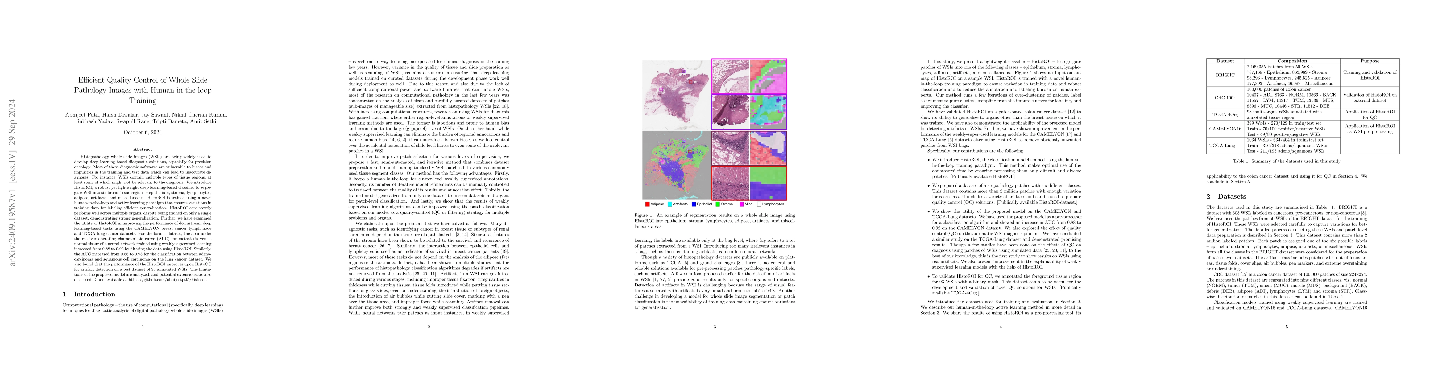
Histopathology whole slide images (WSIs) are being widely used to develop deep learning-based diagnostic solutions, especially for precision oncology. Most of these diagnostic softwares are vulnerable to biases and impurities in the training and test data which can lead to inaccurate diagnoses. For instance, WSIs contain multiple types of tissue regions, at least some of which might not be relevant to the diagnosis. We introduce HistoROI, a robust yet lightweight deep learning-based classifier to segregate WSI into six broad tissue regions -- epithelium, stroma, lymphocytes, adipose, artifacts, and miscellaneous. HistoROI is trained using a novel human-in-the-loop and active learning paradigm that ensures variations in training data for labeling-efficient generalization. HistoROI consistently performs well across multiple organs, despite being trained on only a single dataset, demonstrating strong generalization. Further, we have examined the utility of HistoROI in improving the performance of downstream deep learning-based tasks using the CAMELYON breast cancer lymph node and TCGA lung cancer datasets. For the former dataset, the area under the receiver operating characteristic curve (AUC) for metastasis versus normal tissue of a neural network trained using weakly supervised learning increased from 0.88 to 0.92 by filtering the data using HistoROI. Similarly, the AUC increased from 0.88 to 0.93 for the classification between adenocarcinoma and squamous cell carcinoma on the lung cancer dataset. We also found that the performance of the HistoROI improves upon HistoQC for artifact detection on a test dataset of 93 annotated WSIs. The limitations of the proposed model are analyzed, and potential extensions are also discussed.

AI Key Findings

Get AI-generated insights about this paper's methodology, results, and significance.

Paper Details

PDF Preview

Download PDF

Key Terms

No key terms available for this paper.

Title Authors Year Actions
Scalable Whole Slide Image Representation Using K-Means Clustering and Fisher Vector Aggregation
Ravi Kant Gupta, Shounak Das, Ardhendu Sekhar, Amit Sethi
2025
View Paper
Exploring Self-Supervised Learning for Disease Detection and Classification in Digital Pathology: A review
Abdulahi Mahammed Adem, Ravi Kant, Sonia S, Karan Kumar, Vikas Mittal, Pankaj Jain, Kapil Joshi
2025
View Paper
GrandQC: A comprehensive solution to quality control problem in digital pathology
Zhilong Weng, Alexander Seper, Alexey Pryalukhin, Fabian Mairinger, Claudia Wickenhauser, Marcus Bauer, Lennert Glamann, Hendrik Bläker, Thomas Lingscheidt, Wolfgang Hulla, D. Jonigk, Simon Schallenberg, Andrey Bychkov, Junya Fukuoka, Martin Braun, B. Schömig-Markiefka, Sebastian Klein, Andreas Thiel, Katarzyna Bożek, George J Netto, A. Quaas, Reinhard Buettner, Yuri Tolkach
2024
View Paper
Artificial intelligence in surgical pathology – where do we stand, where do we go?
Chen Sagiv, Ofir Hadar, Abderrahman Najjar, Jens Pahnke
2024
View Paper
Fully Automatic Content-Aware Tiling Pipeline for Pathology Whole Slide Images
Falah Jabar, L. Busund, B. Ricciuti, M. Tafavvoghi, Mette Pøhl, S. Andersen, T. Dønnem, David J. Kwiatkowski, M. Rakaee
2024
View Paper
Classification and Morphological Analysis of Dlbcl Subtypes in H&E-Stained Slides
Ravi Kant Gupta, Mohit Jindal, Garima Jain, E. Sridhar, Subhash Yadav, H. Jain, T. Shet, Uma M. Sakhdeo, M. Sengar, L. Nayak, B. Bagal, Umesh Apkare, Amit Sethi
2024
View Paper
Clustered Patch Embeddings for Permutation-Invariant Classification of Whole Slide Images
Ravi Kant Gupta, Shounak Das, Amit Sethi
2024
View Paper
Efficient Whole Slide Image Classification Through Fisher Vector Representation
Ravi Kant Gupta, Dadi Dharani, Shambhavi Shanker, Amit Sethi
2024
View Paper
Evaluation Metric for Quality Control and Generative Models in Histopathology Images
Pranav Jeevan, Neeraj Nixon, Abhijeet Patil, Amit Sethi
2024
View Paper
Semantic Segmentation Based Quality Control of Histopathology Whole Slide Images
Abhijeet Patil, Garima Jain, Harsh Diwakar, Jay Sawant, Tripti Bameta, Swapnil Rane, Amit Sethi
2024
View Paper
HER2 and Fish Status Prediction in Breast Biopsy H&E-Stained Images Using Deep Learning
Ardhendu Sekhar, Vrinda Goel, Garima Jain, Abhijeet Patil, Ravi Kant Gupta, Amit Sethi
2024
View Paper
Multi-Artifact Detection and Filtering in Digital Pathology Using Intrinsic Image Properties
Varun Kanwar, Addie Woicik, B. Dulken, C. Probert, Z. McCaw
2024
View Paper
Computational methods for metastasis detection in lymph nodes and characterization of the metastasis-free lymph node microarchitecture: A systematic-narrative hybrid review
E. Budginaite, D. R. Magee, M. Kloft, Henry C. Woodruff, H. I. Grabsch
2024
View Paper
iQC: machine-learning-driven prediction of surgical procedure uncovers systematic confounds of cancer whole slide images in specific medical centers
A. J. Schaumberg, M. S. Lewis, R. Nazarian, A. Wadhwa, N. Kane, G. Turner, P. Karnam, P. Devineni, N. Wolfe, R. Kintner, M. Rettig, B. S. Knudsen, I. Garraway, S. Pyarajan
2023
View Paper
Deep Learning for Lung Cancer Diagnosis, Prognosis and Prediction Using Histological and Cytological Images: A Systematic Review
Athena S Davri, Effrosyni Birbas, Theofilos Kanavos, Georgios Ntritsos, N. Giannakeas, A. Tzallas, A. Batistatou
2023
View Paper

Comments (0)

This website uses cookies to ensure you get the best experience. Learn more