ArxivLens

Quick Summary:

This paper introduces a new Quantitative Hyperspectral Image Unmixing (Q-HIU) method for analyzing large-scale Raman micro-spectroscopy data. The method involves SVD-ADC for noise reduction, BGF for background subtraction, and Q-US/PS-NMF for retrieving non-negative spatial concentration maps and spectral profiles, demonstrating its effectiveness on human atherosclerotic aortic tissues.

Journal Reference: Analytica Chimica Acta 1050 (2019) 32-43
DOI: 10.1016/j.aca.2018.11.018
arXiv Id: 1803.01823
Date Published: 2019-03-12
Date Updated: 2019-03-12

Categories:

Key Terms (TF-IDF):

raman (0.277)
non negative (0.253)
micro (0.214)
ps (0.181)
negative (0.171)
spectroscopy (0.167)
image (0.158)
partially (0.149)
concentration (0.149)
supervised (0.135)
chemical (0.132)
quantitative (0.132)
samples (0.128)
method (0.125)
large scale (0.123)
non (0.116)
images (0.114)
spatial (0.114)
efficient (0.105)
alternatively (0.105)
License: http://arxiv.org/licenses/nonexclusive-distrib/1.0/

Summary:

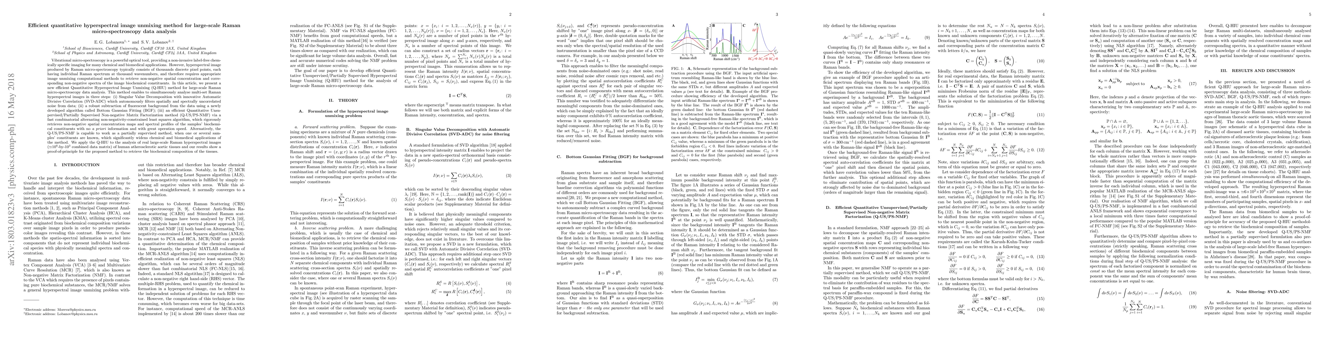
Vibrational micro-spectroscopy is a powerful optical tool, providing a non-invasive label-free chemically specific imaging for many chemical and biomedical applications. However, hyperspectral image produced by Raman micro-spectroscopy typically consists of thousands discrete pixel points, each having individual Raman spectrum at thousand wavenumbers, and therefore requires appropriate image unmixing computational methods to retrieve non-negative spatial concentration and corresponding non-negative spectra of the image biochemical constituents. In this article, we present a new efficient Quantitative Hyperspectral Image Unmixing (Q-HIU) method for large-scale Raman micro-spectroscopy data analysis. This method enables to simultaneously analyse multi-set Raman hyperspectral images in three steps: (i) Singular Value Decomposition with innovative Automatic Divisive Correlation (SVD-ADC) which autonomously filters spatially and spectrally uncorrelated noise from data; (ii) a robust subtraction of fluorescent background from the data using a newly developed algorithm called Bottom Gaussian Fitting (BGF); (iii) an efficient Quantitative Unsupervised/Partially Supervised Non-negative Matrix Factorization method (Q-US/PS-NMF), which rigorously retrieves non-negative spatial concentration maps and spectral profiles of the samples' biochemical constituents with no a priori information and with great operation speed. Alternatively, the Q-US/PS-NMF is capable to work as a partially supervised method, when one or several samples' constituents are known, which significantly widens chemical and biomedical applications of the method. We apply the Q-HIU to the analysis of real large-scale Raman hyperspectral images of human atherosclerotic aortic tissues and our results show a proof-of-principle for the proposed method to retrieve the biochemical composition of the tissues.

Citation & Reference Network Visualization

This graph shows how this paper is connected to other research. The current paper is shown in dark gray, papers that cite it are in green, and papers it references are in blue. You can:

Current Paper
Papers citing this work (Citations)
Papers cited by this work (References)

• Hover over any node to see the paper title

• Click a node to view that paper's details

• Drag nodes to rearrange the graph

• Use the zoom controls (bottom right) to zoom in/out

Citations and References

Title Authors Year Actions
Surface-Enhanced Raman Spectroscopy for Biomedical Applications: Recent Advances and Future Challenges.
Linley Li Lin, Ramon A. Alvarez‐Puebla, Luis M. Liz-Marzán, M. Trau, Jing Wang, Laura Fabris, Xiang Wang, Guo-kun Liu, Shuping Xu, X. Han, Liangbao Yang, Aiguo Shen, Shikuan Yang, Yikai Xu, Chunchun Li, Jinqing Huang, Shaoying Liu, Jian-An Huang, I. Srivastava, Ming Li, Limei Tian, Lam Bang Thanh Nguyen, Xinyuan Bi, D. Cialla‐May, P. Matousek, Nicholas Stone, Randy P. Carney, Wei Ji, Wei Song, Zhou Chen, I. Phang, Malou Henriksen-Lacey, Haoran Chen, Zongyu Wu, Heng Guo, Hao Ma, Gennadii Ustinov, Siheng Luo, Sara Mosca, B. Gardner, Yi-Tao Long, Juergen Popp, B. Ren, Shuming Nie, Binggen Zhao, X. Ling, Jian Ye
2025
View Paper
Hyperspectral Raman imaging with multivariate curve resolution-alternating least square (MCR-ALS) analysis for xylazine-containing drug mixtures.
Xuyang He
2024
View Paper
Hybrid brain tumor classification of histopathology hyperspectral images by linear unmixing and an ensemble of deep neural networks
I. A. Cruz-Guerrero, D. U. Campos-Delgado, A. Mejía-Rodríguez, Raquel León, S. Ortega, H. Fabelo, R. Camacho, M. L. Plaza, G. Callicó
2024
View Paper
Assessment of unmixing approaches for the quantitation of SERS nanoparticles in highly multiplexed spectral images.
Alexander T Czaja, Samer Awad, O. Eremina, A. Fernando, Cristina Zavaleta
2024
View Paper
Nondestructive 3D imaging and quantification of hydrated biofilm matrix by confocal Raman microscopy coupled with non-negative matrix factorization.
Xiao-Yang Liu, Shuxia Guo, T. Bocklitz, P. Rösch, J. Popp, Hanqing Yu
2021
View Paper
Fast denoising and lossless spectrum extraction in stimulated Raman scattering microscopy
Yanping Li, Binglin Shen, Gengjin Zou, Shiqi Wang, J. Qu, Rui Hu, Liwei Liu
2021
View Paper
Simultaneous In Situ Characterizations of Ultrashort Laser Pulses and the Nonlinear Susceptibility of the Irradiated Medium via Time-Resolved Hybrid Coherent Anti-Stokes Raman Scattering Spectroscopy.
Tao Cao, Zhou Li, Jikun Yan, Ziyue Guo, Shaozhen Liu, K. Hu, Ruxin Li, A. Sokolov, Jiahui Peng
2021
View Paper
Imaging of PD-L1 in single cancer cells by SERS-based hyperspectral analysis.
Wei Zhang, Jake S. Rhodes, Kevin R. Moon, B. Knudsen, Linda Nokolova, Anhong Zhou
2020
View Paper
debCAM: a bioconductor R package for fully unsupervised deconvolution of complex tissues
Lulu Chen, Chiung-Ting Wu, Niya Wang, D. Herrington, R. Clarke, Y. Wang
2020
View Paper
SVD-clustering, a general image-analyzing method explained and demonstrated on model and Raman micro-spectroscopic maps
B. Szalontai, M. Debreczeny, K. Fintor, C. Bagyinka
2020
View Paper
Inhomogeneous Molecular Distributions and Cytochrome Types and Redox States in Fungal Cells Revealed by Raman Hyperspectral Imaging Using Multivariate Curve Resolution-Alternating Least Squares.
Mitsuru Yasuda, Norio Takeshita, Shinsuke Shigeto
2019
View Paper

Similar Papers

Hyperspectral unmixing for Raman spectroscopy via physics-constrained autoencoders

Mauricio Barahona, Georgios Papadopoulos, Dimitar Georgiev, Álvaro Fernández-Galiana, Simon Vilms Pedersen, Ruoxiao Xie, Molly M. Stevens

Published: 2024-03-08

Transformer for Multitemporal Hyperspectral Image Unmixing

Tao Li, Hang Li, Zhenwei Shi, Qiankun Dong, Xueshuo Xie, Xia Xu

Published: 2024-07-15

Comments (0)

This website uses cookies to ensure you get the best experience. Learn more