Efficient Transformer-Based Localized Patch Sampling for Choroid Plexus Segmentation in Multiple Sclerosis
Publication
Metrics
AI Quick Summary
A SwinUNETR-based pipeline using targeted intra- and peri-ventricular patch sampling accurately segments the lateral ventricle choroid plexus (LVCP) in multiple sclerosis MRI, outperforming the UXNET model and remaining robust with standalone and multi-modal inputs. It achieves high Dice scores (≈0.868 with MPRAGE+FLAIR) while dramatically reducing computational cost (≈99% lower GFLOPs), enabling scalable, efficient LVCP analysis in clinical and research settings.
Paper Preview
Abstract
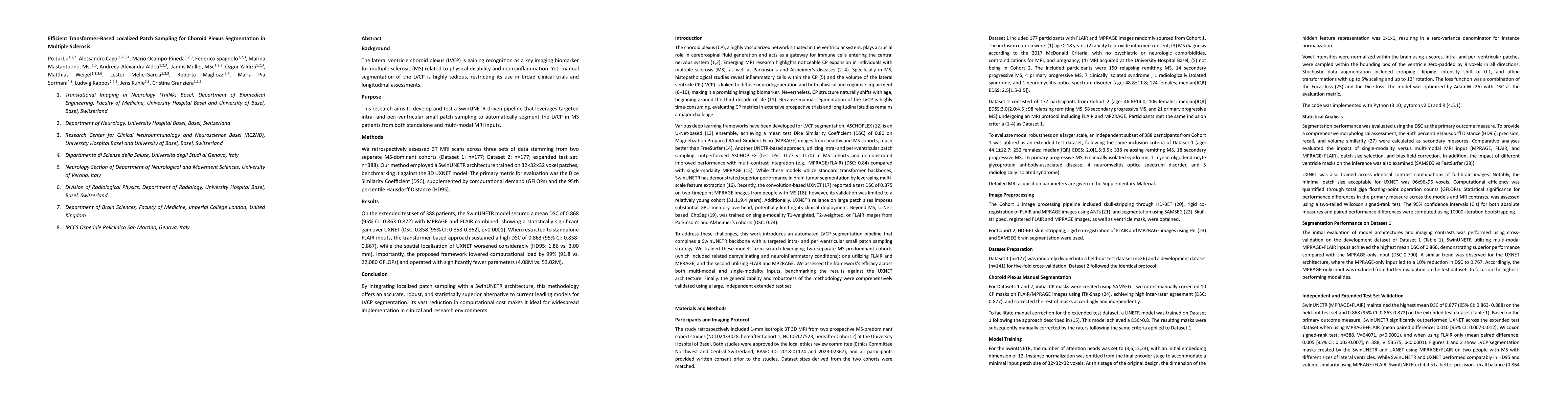
Background: The lateral ventricle choroid plexus (LVCP) is gaining recognition as a key imaging biomarker for multiple sclerosis (MS) related to physical disability and neuroinflammation. Yet, manual segmentation of the LVCP is highly tedious, restricting its use in broad clinical trials and longitudinal assessments. This research aims to develop a SwinUNETR-driven pipeline that leverages targeted intra- and peri-ventricular small patch sampling to automatically segment the LVCP in MS from both standalone and multi-modal MRI inputs. Methods: We retrospectively assessed 3T MRI scans across three sets of data stemming from two separate MS-dominant cohorts (Dataset 1: n=177; Dataset 2: n=177; expanded test set: n=388). Our method employed a SwinUNETR architecture trained on 32x32x32 voxel patches, benchmarking it against the 3D UXNET model. The primary metric for evaluation was the Dice Similarity Coefficient (DSC), supplemented by computational demand (GFLOPs) and the 95th percentile Hausdorff Distance (HD95). Results: On the extended test set, the SwinUNETR model secured a mean DSC of 0.868 (95% CI: 0.863-0.872) with MPRAGE and FLAIR combined, showing a statistically significant gain over UXNET (DSC: 0.858 [95% CI: 0.853-0.862], p<0.0001). When restricted to standalone FLAIR inputs, the transformer-based approach sustained a high DSC of 0.863, while the spatial localization of UXNET worsened considerably (HD95: 1.86 vs. 3.00 mm). Importantly, the proposed framework lowered computational load by 99% (91.8 vs. 22,080 GFLOPs). By integrating localized patch sampling with a SwinUNETR architecture, this methodology offers an accurate, robust, and statistically superior alternative to current leading models for LVCP segmentation. Its vast reduction in computational cost makes it ideal for widespread implementation in clinical and research environments.
AI Key Findings
Get AI-generated insights about this paper's methodology, results, significance, and more — seven facets brought into focus.
Discussion 0