Summary
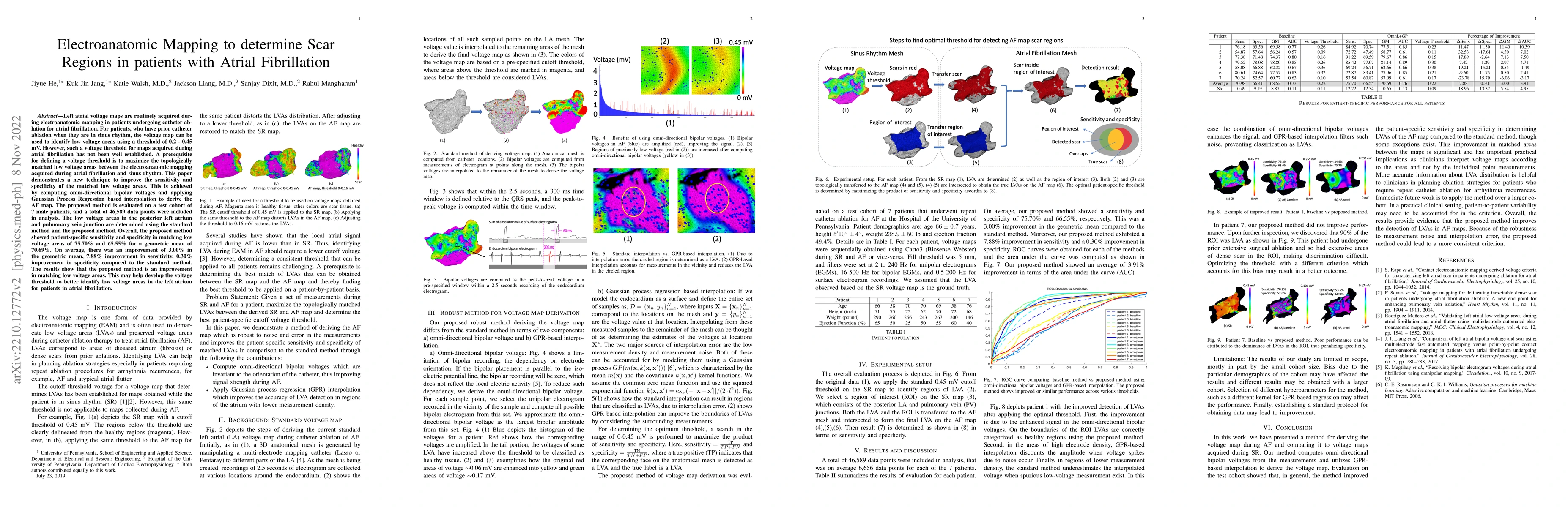
Left atrial voltage maps are routinely acquired during electroanatomic mapping in patients undergoing catheter ablation for atrial fibrillation. For patients, who have prior catheter ablation when they are in sinus rhythm, the voltage map can be used to identify low voltage areas using a threshold of 0.2 - 0.45 mV. However, such a voltage threshold for maps acquired during atrial fibrillation has not been well established. A prerequisite for defining a voltage threshold is to maximize the topologically matched low voltage areas between the electroanatomic mapping acquired during atrial fibrillation and sinus rhythm. This paper demonstrates a new technique to improve the sensitivity and specificity of the matched low voltage areas. This is achieved by computing omni-directional bipolar voltages and applying Gaussian Process Regression based interpolation to derive the atrial fibrillation map. The proposed method is evaluated on a test cohort of 7 male patients, and a total of 46,589 data points were included in analysis. The low voltage areas in the posterior left atrium and pulmonary vein junction are determined using the standard method and the proposed method. Overall, the proposed method showed patient-specific sensitivity and specificity in matching low voltage areas of 75.70% and 65.55% for a geometric mean of 70.69%. On average, there was an improvement of 3.00% in the geometric mean, 7.88% improvement in sensitivity, 0.30% improvement in specificity compared to the standard method. The results show that the proposed method is an improvement in matching low voltage areas. This may help develop the voltage threshold to better identify low voltage areas in the left atrium for patients in atrial fibrillation.
AI Key Findings
Get AI-generated insights about this paper's methodology, results, and significance.
Paper Details
PDF Preview
Key Terms
Citation Network
Current paper (gray), citations (green), references (blue)
Display is limited for performance on very large graphs.
Similar Papers
Found 4 papersAI-guided spatiotemporal dispersion mapping for individualized ablation in an all-comer cohort with atrial fibrillation.
Hindricks, Gerhard, Heil, Emanuel, Dagres, Nikolaos et al.
Integrating Deep Learning in Cardiology: A Comprehensive Review of Atrial Fibrillation, Left Atrial Scar Segmentation, and the Frontiers of State-of-the-Art Techniques
Malitha Gunawardhana, Jichao Zhao, Anuradha Kulathilaka
Multi-Depth Boundary-Aware Left Atrial Scar Segmentation Network
Mengjun Wu, Wangbin Ding, Liqin Huang et al.
| Title | Authors | Year | Actions |
|---|
Comments (0)