ArxivLens

Quick Summary:

This paper proposes a new spectroscopic technique called electron- and light-induced stimulated Raman (ELISR) scattering, which combines electron microscopy's high spatial resolution with Raman spectroscopy's molecular sensitivity. The technique uses electron-beam excitation of plasmonic nanoparticles to achieve significant stimulated Raman enhancement.

Journal Reference: Phys. Rev. B 102, 085406 (2020)
DOI: 10.1103/PhysRevB.102.085406
arXiv Id: 2001.11638
Comments: 7 pages, 4 figures, Amr A. E. Saleh and Daniel K. Angell contributed equally to this work
Date Published: 2020-08-12
Date Updated: 2020-08-12

Key Terms (TF-IDF):

raman (0.320)
microscopy (0.277)
vibrational (0.248)
spectroscopy (0.240)
electron (0.239)
diffraction (0.187)
geometries (0.182)
resolution (0.161)
beam (0.160)
excitation (0.156)
limited (0.139)
spatial (0.132)
energy loss (0.127)
nanoparticles (0.121)
light (0.121)
termed (0.119)
broadband (0.115)
promise (0.114)
stokes (0.113)
importantly (0.110)
License: http://arxiv.org/licenses/nonexclusive-distrib/1.0/

Summary:

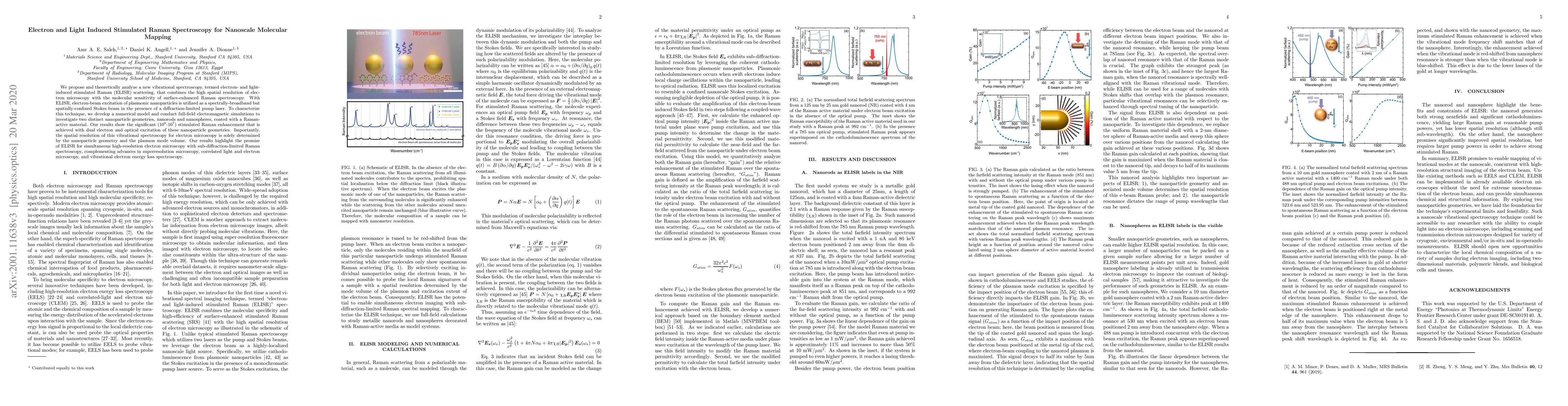
We propose and theoretically analyze a new vibrational spectroscopy, termed electron- and light-induced stimulated Raman (ELISR) scattering, that combines the high spatial resolution of electron microscopy with the molecular sensitivity of surface-enhanced Raman spectroscopy. With ELISR, electron-beam excitation of plasmonic nanoparticles is utilized as a spectrally-broadband but spatially-confined Stokes beam in the presence of a diffraction-limited pump laser. To characterize this technique, we develop a numerical model and conduct full-field electromagnetic simulations to investigate two distinct nanoparticle geometries, nanorods and nanospheres, coated with a Raman-active material. Our results show the significant ($10^6$-$10^7$) stimulated Raman enhancement that is achieved with dual electron and optical excitation of these nanoparticle geometries. Importantly, the spatial resolution of this vibrational spectroscopy for electron microscopy is solely determined by the nanoparticle geometry and the plasmon mode volume. Our results highlight the promise of ELISR for simultaneous high-resolution electron microscopy with sub-diffraction-limited Raman spectroscopy, complementing advances in superresolution microscopy, correlated light and electron microscopy, and vibrational electron energy loss spectroscopy.

Citation & Reference Network Visualization

This graph shows how this paper is connected to other research. The current paper is shown in dark gray, papers that cite it are in green, and papers it references are in blue. You can:

Current Paper
Papers citing this work (Citations)
Papers cited by this work (References)

• Hover over any node to see the paper title

• Click a node to view that paper's details

• Drag nodes to rearrange the graph

• Use the zoom controls (bottom right) to zoom in/out

Citations and References

Title Authors Year Actions
Nanoscale and ultrafast in situ techniques to probe plasmon photocatalysis
Claire C. Carlin, Alan X. Dai, Alexander Al-Zubeidi, Emma M. Simmerman, Hyuncheol Oh, Niklas Gross, Stephen A. Lee, Stephan Link, C. Landes, Felipe H. da Jornada, Jennifer A. Dionne
2023
View Paper
The Light Years: Combined optical and environmental electron microscopy to visualize photonic processes with atomic-scale resolution
J. Dionne, F. Hayee, Michal Vadai, D. Angell, Amr A. E. Saleh, Katherine Sytwu
2019
View Paper

Comments (0)

This website uses cookies to ensure you get the best experience. Learn more