Summary
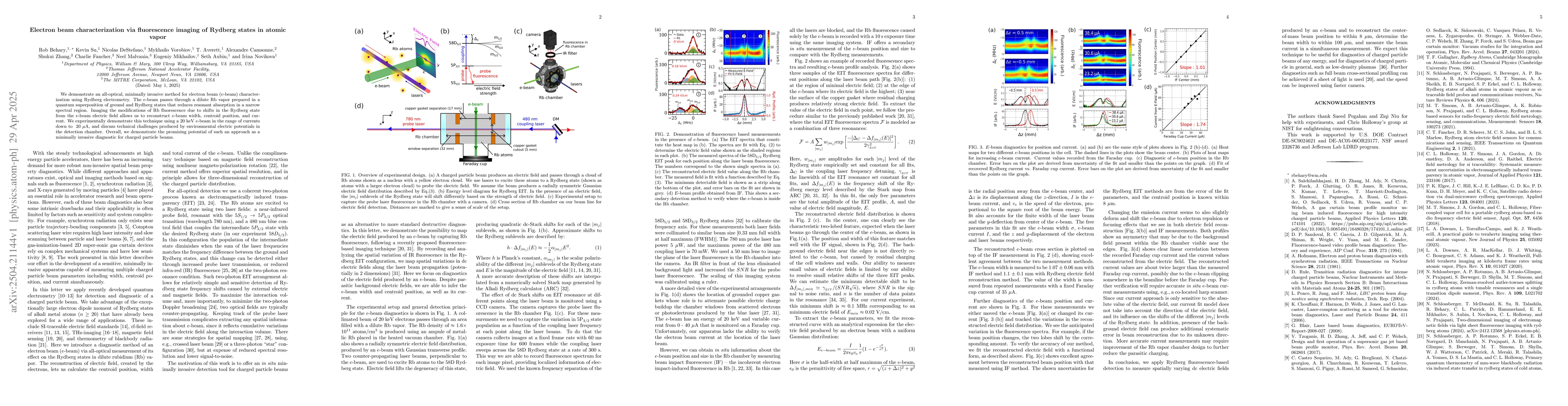
We demonstrate an all-optical, minimally invasive method for electron beam (e-beam) characterization using Rydberg electrometry. The e-beam passes through a dilute Rb vapor prepared in a quantum superposition of ground and Rydberg states that reduces resonant absorption in a narrow spectral region. Imaging the modifications of Rb fluorescence due to shifts in the Rydberg state from the e-beam electric field allows us to reconstruct e-beam width, centroid position, and current. We experimentally demonstrate this technique using a 20 keV e-beam in the range of currents down to 20 $\mu$A, and discuss technical challenges produced by environmental electric potentials in the detection chamber. Overall, we demonstrate the promising potential of such an approach as a minimally invasive diagnostic for charged particle beams.
AI Key Findings
Generated Jun 08, 2025
Methodology
The research employs an all-optical, minimally invasive method for electron beam characterization using Rydberg electrometry. A dilute Rb vapor is prepared in a quantum superposition of ground and Rydberg states, which reduces resonant absorption in a narrow spectral region. The modifications of Rb fluorescence due to shifts in the Rydberg state from the e-beam electric field are imaged to reconstruct e-beam width, centroid position, and current.
Key Results
- Demonstrated characterization of a 20 keV electron beam with currents as low as 20 μA.
- Successfully imaged modifications of Rb fluorescence to extract e-beam width, centroid position, and current.
- Identified technical challenges posed by environmental electric potentials in the detection chamber.
Significance
This method offers a minimally invasive diagnostic for charged particle beams, which could be crucial for applications requiring precise beam characterization without disturbance.
Technical Contribution
Introduction of Rydberg electrometry for all-optical, minimally invasive electron beam characterization.
Novelty
This work presents a novel approach utilizing Rydberg states in atomic vapor for e-beam characterization, distinct from conventional methods that often involve direct interaction or significant perturbation of the beam.
Limitations
- Technical challenges due to environmental electric potentials in the detection chamber.
- Limited to specific experimental conditions (e.g., 20 keV e-beam, dilute Rb vapor)
Future Work
- Explore applicability to different types and energies of electron beams.
- Develop strategies to mitigate the impact of environmental electric potentials
Paper Details
PDF Preview
Citation Network
Current paper (gray), citations (green), references (blue)
Display is limited for performance on very large graphs.
Similar Papers
Found 4 papersTwo-dimensional imaging of electromagnetic fields via light sheet fluorescence imaging with Rydberg atoms
Nikunjkumar Prajapati, Christopher L. Holloway, Daniel Hammerland et al.
Imaging of induced surface charge distribution effects in glass vapor cells used for Rydberg atom-based sensors
Nikunjkumar Prajapati, Samuel Berweger, Christopher L. Holloway et al.
No citations found for this paper.
Comments (0)