Electron microscopy of microwave-synthesized rare-earth chromites
Publication
Metrics
AI Quick Summary
This study synthesizes rare-earth chromites using microwave techniques and employs X-ray diffraction, HRTEM, and FE-SEM to confirm the perovskite crystal structure and particle sizes. Magnetization measurements correlate with structural data, validating the synthesis method.
Paper Preview
Abstract
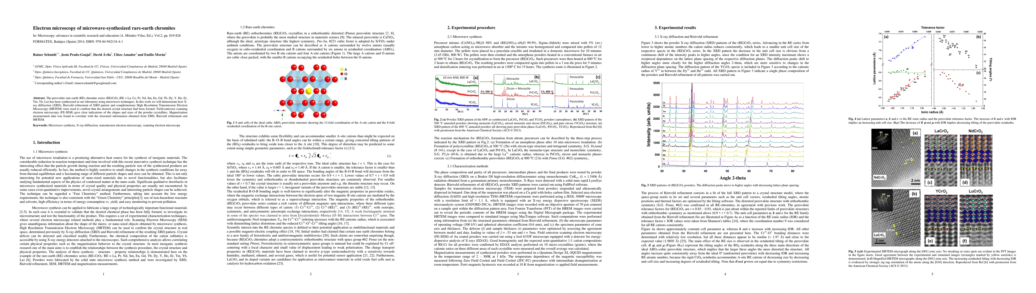
The perovskite rare-earth (RE) chromite series (RE)CrO3 (RE = La, Ce, Pr, Nd, Sm, Eu, Gd, Tb, Dy, Y, Ho, Er, Tm, Yb, Lu) has been synthesized in our laboratory using microwave techniques. In this work we will demonstrate how X-ray diffraction (XRD), Rietveld refinement of XRD pattern and complementary High Resolution Transmission Electron Microscopy (HRTEM) were used to confirm that the desired crystal structure had been formed. Field-emission scanning electron microscopy (FE-SEM) gave clear indications of the shapes and sizes of the powder crystallites. Magnetization measurement data was found to correlate with the structural information obtained from XRD, Rietveld refinement and HRTEM.
AI Key Findings
Get AI-generated insights about this paper's methodology, results, significance, and more — seven facets brought into focus.
Impact
Paper Details
PDF Preview
Key Terms
Citation Network
Current paper (gray), citations (green), references (blue)
Display is limited for performance on very large graphs.
Discussion 0