Electron Spin Resonance Spectroscopy via Relaxation of Solid-State Spin Probes at the Nanoscale
Publication
Metrics
AI Quick Summary
This paper introduces a novel method for nanoscale Electron Spin Resonance (ESR) spectroscopy by measuring the relaxation time of a single-spin probe in solid-state systems, demonstrating high spatial resolution and agreement with theoretical predictions using nitrogen-vacancy centres in diamond. The technique promises to enable ESR spectroscopy at the atomic and molecular scale.
Paper Preview
Abstract
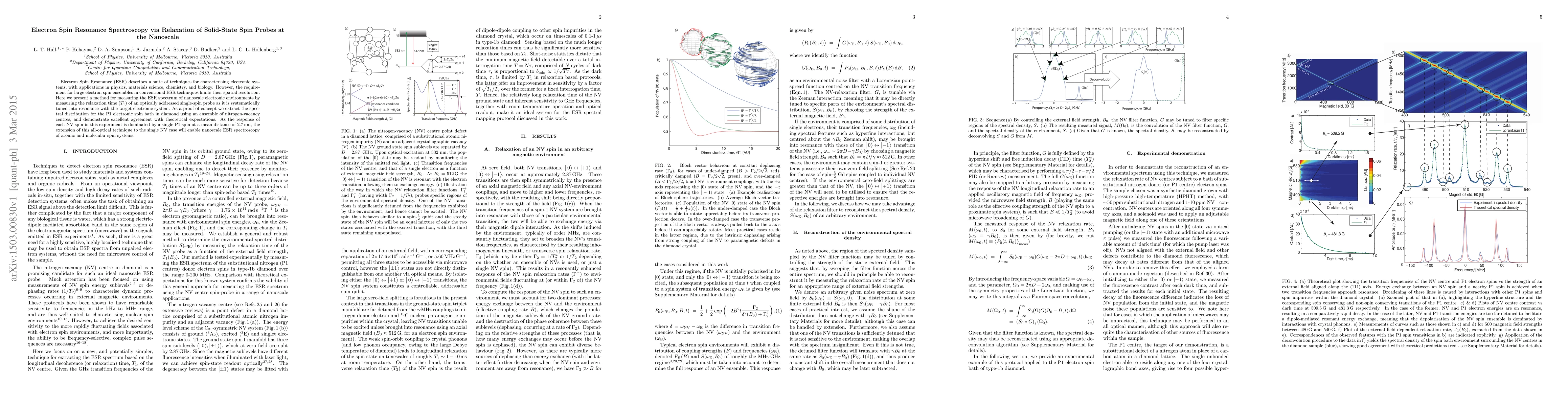
Electron Spin Resonance (ESR) describes a suite of techniques for characterising electronic systems, with applications in physics, materials science, chemistry, and biology. However, the requirement for large electron spin ensembles in conventional ESR techniques limits their spatial resolution. Here we present a method for measuring the ESR spectrum of nanoscale electronic environments by measuring the relaxation time ($T_1$) of an optically addressed single-spin probe as it is systematically tuned into resonance with the target electronic system. As a proof of concept we extract the spectral distribution for the P1 electronic spin bath in diamond using an ensemble of nitrogen-vacancy centres, and demonstrate excellent agreement with theoretical expectations. As the response of each NV spin in this experiment is dominated by a single P1 spin at a mean distance of 2.7\,nm, the extension of this all-optical technique to the single NV case will enable nanoscale ESR spectroscopy of atomic and molecular spin systems.
AI Key Findings
Get AI-generated insights about this paper's methodology, results, significance, and more — seven facets brought into focus.
Impact
Paper Details
PDF Preview
Key Terms
Citation Network
Current paper (gray), citations (green), references (blue)
Display is limited for performance on very large graphs.
Discussion 0