Publication
Metrics
AI Quick Summary
This study employs low-temperature X-ray photoemission electron microscopy (X-PEEM) to map the electrostatic potential at domain walls in ferroelectric Er0.99Ca0.01MnO3, revealing uncompensated bound charges. This method provides superior resolution and faster data acquisition compared to low-temperature electrostatic force microscopy (EFM).
Paper Preview
Abstract
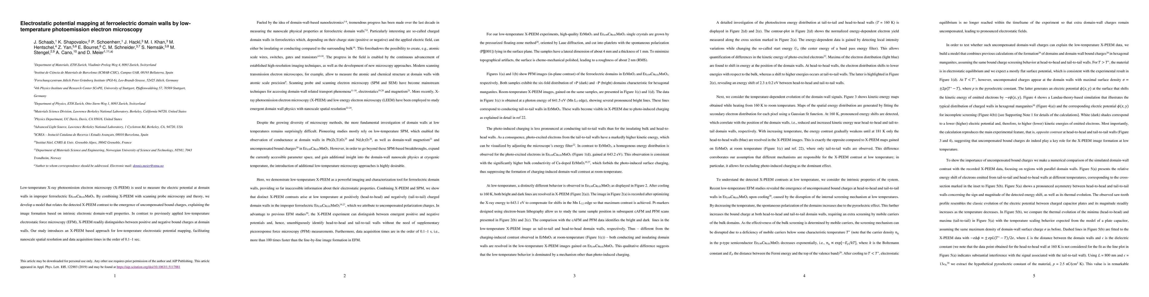
Low-temperature X-ray photoemission electron microscopy (X-PEEM) is used to measure the electric potential at domain walls in improper ferroelectric Er0.99Ca0.01MnO3. By combining X-PEEM with scanning probe microscopy and theory, we develop a model that relates the detected X-PEEM contrast to the emergence of uncompensated bound charges, explaining the image formation based on intrinsic electronic domain-wall properties. In contrast to previously applied low-temperature electrostatic force microscopy (EFM), X-PEEM readily distinguishes between positive and negative bound charges at domain walls. Our study introduces an X-PEEM based approach for low-temperature electrostatic potential mapping, facilitating nanoscale spatial resolution and data acquisition times in the order of 0.1-1 sec.
AI Key Findings
Get AI-generated insights about this paper's methodology, results, significance, and more — seven facets brought into focus.
Impact
Paper Details
Authors
PDF Preview
Key Terms
Citation Network
Current paper (gray), citations (green), references (blue)
Display is limited for performance on very large graphs.
Discussion 0