ArxivLens

Publication

Published: Sep 15, 2025
Categories: cs.CV

Metrics

Source: ArXiv

Quick Answers

What methodology did the authors use?

The research proposes TetHeart, a unified framework for reconstructing heart motion from either full stacks of 2D MRI slices or sparse sets down to a single slice. It combines 3D shape reconstruction with motion tracking using deep learning techniques, including a novel AFA module for handling sparse 2D inputs and spatial information integration. More in Methodology →

What are the key results?

TetHeart successfully infers full 3D heart shape and motion from sparse real-time CMR slices for the first time. — The framework demonstrates superior performance compared to existing methods in both full-stack and sparse input scenarios. More in Key Results →

Why is this work significant?

This research enables real-time, image-guided interventions in cardiology by allowing accurate heart motion reconstruction from limited MRI data. It bridges the gap between retrospective analysis and live clinical use, paving the way for digital twins of beating hearts in medical applications. More in Significance →

What are the main limitations?

The method relies on high-quality 2D slice inputs which may not always be available in real-time clinical settings. — Performance may degrade with extremely sparse inputs beyond a single slice without additional data augmentation strategies. More in Limitations →

Paper Preview

Abstract

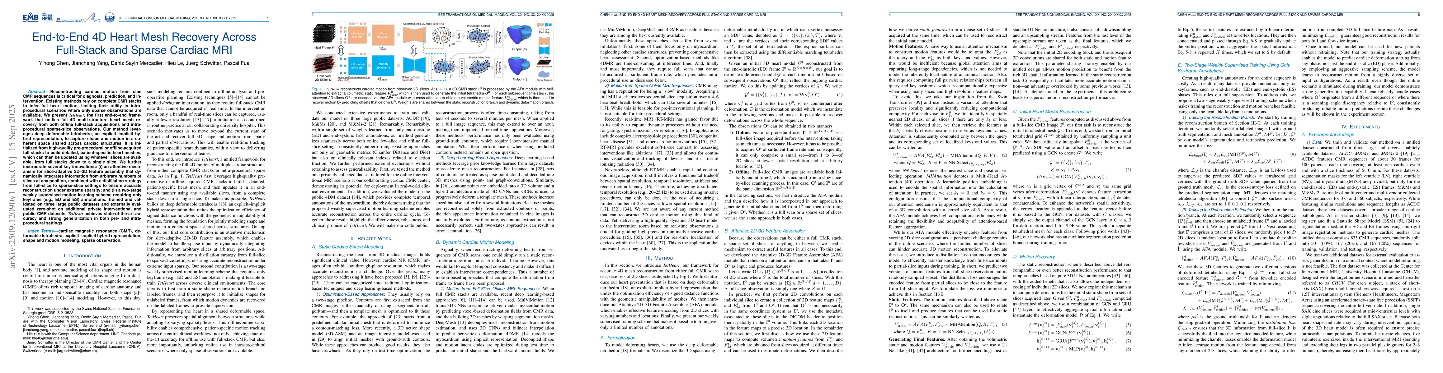
Reconstructing cardiac motion from cine CMR sequences is critical for diagnosis, prediction, and intervention. Existing methods rely on complete CMR stacks to infer full heart motion, limiting their utility in intra-procedural scenarios where only sparse observations are available. We present TetHeart, the first end-to-end framework that unifies full 4D multi-structure heart mesh recovery from both offline full-stack acquisitions and intra-procedural sparse-slice observations. Our method leverages deep deformable tetrahedra, an explicit-implicit hybrid representation, to capture shape and motion in a coherent space shared across cardiac structures. It is initialized from high-quality pre-procedural or offline-acquired full stacks to build detailed, patient-specific heart meshes, which can then be updated using whatever slices are available, from full stacks down to a single slice. We further incorporate several key innovations: (i) an attentive mechanism for slice-adaptive 2D-3D feature assembly that dynamically integrates information from arbitrary numbers of slices at any position, combined with a distillation strategy from full-slice to sparse-slice settings to ensure accurate reconstruction under extreme sparsity; and (ii) a two-stage weakly supervised motion learning scheme requiring only keyframe (e.g., ED and ES) annotations. Trained and validated on three large public datasets and externally evaluated zero-shot on additional private interventional and public CMR datasets, TetHeart achieves state-of-the-art accuracy and strong generalization in both pre- and intra-procedural settings.

Key Findings, in focus

Seven facets of this paper, analysed and brought into focus by AI.

AI-generated · verify against source Generated Oct 02, 2025
In focus · one-line synthesis

This research enables real-time, image-guided interventions in cardiology by allowing accurate heart motion reconstruction from limited MRI data.

01
MethodologyHow they did it

The research proposes TetHeart, a unified framework for reconstructing heart motion from either full stacks of 2D MRI slices or sparse sets down to a single slice. It combines 3D shape reconstruction with motion tracking using deep learning techniques, including a novel AFA module for handling sparse 2D inputs and spatial information integration.

02
ResultsWhat they found
  • TetHeart successfully infers full 3D heart shape and motion from sparse real-time CMR slices for the first time.
  • The framework demonstrates superior performance compared to existing methods in both full-stack and sparse input scenarios.
  • The integration of spatial information and parameter sharing between reconstruction and motion branches significantly improves robustness and generalization.
03
SignificanceWhy it matters

This research enables real-time, image-guided interventions in cardiology by allowing accurate heart motion reconstruction from limited MRI data. It bridges the gap between retrospective analysis and live clinical use, paving the way for digital twins of beating hearts in medical applications.

04
Technical ContributionsWhat's new technically

The technical contribution includes the AFA module for handling sparse 2D inputs, spatial position encoding integration in attention mechanisms, and parameter sharing between reconstruction and motion branches to transfer 3D knowledge effectively.

05
NoveltyWhat sets it apart

This work introduces a novel framework that enables full 3D heart motion reconstruction from sparse real-time MRI data, combining shape reconstruction with motion tracking in a unified approach, which is a significant advancement over previous methods that typically handle either shape or motion separately.

06
LimitationsWhat to be cautious about
  • The method relies on high-quality 2D slice inputs which may not always be available in real-time clinical settings.
  • Performance may degrade with extremely sparse inputs beyond a single slice without additional data augmentation strategies.
07
Future WorkWhere it goes next
  • Deployment of TetHeart in real interventional MRI suites for clinical validation.
  • Extending the framework to other moving organs and integrating with complementary imaging modalities.
  • Developing predictive digital twins that can anticipate organ motion under various physiological and intervention conditions.

Paper Details

Paper ID: 2509.12090
Categories:
cs.CV

How to Cite This Paper

Reference manager: RIS EndNote


Discussion 0

This website uses cookies to ensure you get the best experience. Learn more