Endoscopic en-face optical coherence tomography and fluorescence imaging using correlation-based probe tracking
Publication
Metrics
AI Quick Summary
This paper presents a method for transforming a 1D scanning probe in endoscopic optical coherence tomography (OCT) into a 2D scanning system using manual scanning along a second axis. It combines OCT with fluorescence imaging and corrects for probe motion in 3D to provide real-time en-face OCT slices and fluorescence images.
Paper Preview
Abstract
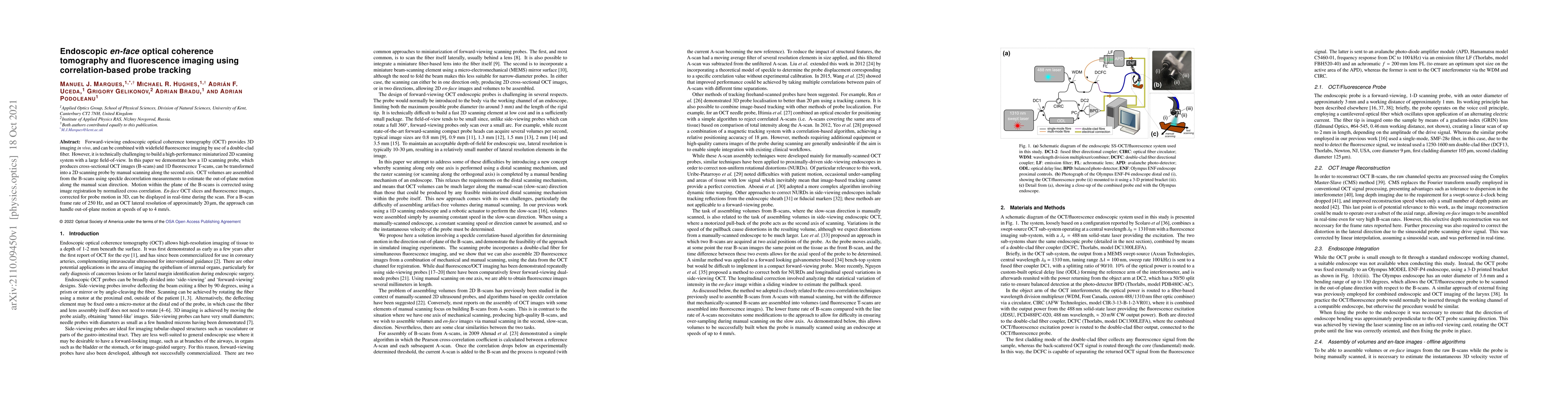
Forward-viewing endoscopic optical coherence tomography (OCT) provides 3D imaging in vivo, and can be combined with widefield fluorescence imaging by use of a double-clad fiber. However, it is technically challenging to build a high-performance miniaturized 2D scanning system with a large field-of-view. In this paper we demonstrate how a 1D scanning probe, which produces cross-sectional OCT images (B-scans) and 1D fluorescence T-scans, can be transformed into a 2D scanning probe by manual scanning along the second axis. OCT volumes are assembled from the B-scans using speckle decorrelation measurements to estimate the out-of-plane motion along the manual scan direction. Motion within the plane of the B-scans is corrected using image registration by normalized cross correlation. En-face OCT slices and fluorescence images, corrected for probe motion in 3D, can be displayed in real-time during the scan. For a B-scan frame rate of 250 Hz, and an OCT lateral resolution of approximately 20 micrometers, the approach can handle out-of-plane motion at speeds of up to 4 mm/s.
AI Key Findings
Get AI-generated insights about this paper's methodology, results, significance, and more — seven facets brought into focus.
Impact
Paper Details
Authors
PDF Preview
Key Terms
Citation Network
Current paper (gray), citations (green), references (blue)
Display is limited for performance on very large graphs.
Discussion 0