Publication

Published: Apr 08, 2026
Categories: cs.CV, cs.AI

Metrics

Source: ArXiv

Quick Actions

View PDF

Quick Answers

What methodology did the authors use?

Patient-specific energy manifolds are learned from a baseline multi‑sequence MRI scan by training an implicit neural representation via denoising score matching in a 5‑dimensional sequence space; the resulting energy function defines tissue regimes as basins of attraction and is used as a fixed geometric reference for longitudinal analysis. More in Methodology →

What are the key results?

Stable disease follow‑ups remain confined to low‑energy basins with no systematic drift toward the tumor basin, indicating no recurrence. — In a recurrence case, voxel energy distributions progressively shift toward the baseline tumor basin and a new tumor basin emerges before radiological evidence, suggesting early detection. More in Key Results →

Why is this work significant?

This framework offers a segmentation‑free, patient‑specific geometric reference for longitudinal mpMRI, potentially enabling earlier detection of tumor recurrence and providing a new paradigm for tissue‑at‑risk tracking in neuro‑oncology. More in Significance →

What are the main limitations?

The study is limited to two pediatric brain tumor cases, serving only as a proof‑of‑concept without clinical validation. — The method relies on accurate registration and manual ROI placement for baseline basin definition, which may introduce variability. More in Limitations →

Paper Preview

Abstract

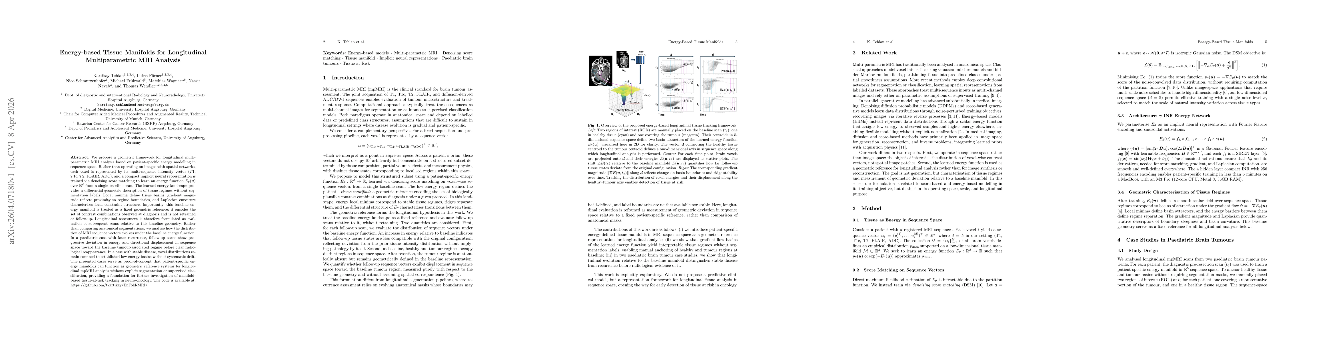
We propose a geometric framework for longitudinal multi-parametric MRI analysis based on patient-specific energy modelling in sequence space. Rather than operating on images with spatial networks, each voxel is represented by its multi-sequence intensity vector ($T1$, $T1c$, $T2$, FLAIR, ADC), and a compact implicit neural representation is trained via denoising score matching to learn an energy function $E_θ(\mathbf{u})$ over $\mathbb{R}^d$ from a single baseline scan. The learned energy landscape provides a differential-geometric description of tissue regimes without segmentation labels. Local minima define tissue basins, gradient magnitude reflects proximity to regime boundaries, and Laplacian curvature characterises local constraint structure. Importantly, this baseline energy manifold is treated as a fixed geometric reference: it encodes the set of contrast combinations observed at diagnosis and is not retrained at follow-up. Longitudinal assessment is therefore formulated as evaluation of subsequent scans relative to this baseline geometry. Rather than comparing anatomical segmentations, we analyse how the distribution of MRI sequence vectors evolves under the baseline energy function. In a paediatric case with later recurrence, follow-up scans show progressive deviation in energy and directional displacement in sequence space toward the baseline tumour-associated regime before clear radiological reappearance. In a case with stable disease, voxel distributions remain confined to established low-energy basins without systematic drift. The presented cases serve as proof-of-concept that patient-specific energy manifolds can function as geometric reference systems for longitudinal mpMRI analysis without explicit segmentation or supervised classification, providing a foundation for further investigation of manifold-based tissue-at-risk tracking in neuro-oncology.

AI Key Findings

Generated Apr 09, 2026

Methodology — What approach did the authors take?

Patient-specific energy manifolds are learned from a baseline multi‑sequence MRI scan by training an implicit neural representation via denoising score matching in a 5‑dimensional sequence space; the resulting energy function defines tissue regimes as basins of attraction and is used as a fixed geometric reference for longitudinal analysis.

Key Results — What are the main findings?

  • Stable disease follow‑ups remain confined to low‑energy basins with no systematic drift toward the tumor basin, indicating no recurrence.
  • In a recurrence case, voxel energy distributions progressively shift toward the baseline tumor basin and a new tumor basin emerges before radiological evidence, suggesting early detection.
  • The baseline energy manifold captures tissue regime geometry without requiring segmentation or supervised labels, enabling continuous monitoring of contrast vector evolution.

Significance — Why does this research matter?

This framework offers a segmentation‑free, patient‑specific geometric reference for longitudinal mpMRI, potentially enabling earlier detection of tumor recurrence and providing a new paradigm for tissue‑at‑risk tracking in neuro‑oncology.

Technical Contribution — What is the technical contribution?

A continuous scalar energy field over voxel‑wise contrast vectors is learned via denoising score matching, providing smooth basins, gradients, and Laplacian curvature that characterize tissue regimes and enable quantitative longitudinal deviation analysis.

Novelty — What is new about this work?

Unlike conventional image‑space segmentation or clustering, this work operates in sequence space, learns a patient‑specific energy manifold without labels, and uses it as a fixed geometric reference for longitudinal monitoring, offering a novel, segmentation‑free approach to early recurrence detection.

Limitations — What are the limitations of this study?

  • The study is limited to two pediatric brain tumor cases, serving only as a proof‑of‑concept without clinical validation.
  • The method relies on accurate registration and manual ROI placement for baseline basin definition, which may introduce variability.

Future Work — What did the authors propose for future work?

  • Validate the approach on larger, diverse cohorts to assess robustness and sensitivity to early recurrence.
  • Integrate spatial information and population‑level comparisons to refine basin definitions and enhance clinical applicability.

Paper Details

Paper ID: 2604.07180
Comments: The code is available at https://github.com/tkartikay/EnFold-MRI
Categories:
cs.CV cs.AI

How to Cite This Paper

BibTeX
@article{tehlan2026energy,
  title         = {Energy-based Tissue Manifolds for Longitudinal Multiparametric MRI Analysis},
  author        = {Tehlan, Kartikay and Förner, Lukas and Wagner, Matthias and others},
  year          = {2026},
  eprint        = {2604.07180},
  archivePrefix = {arXiv},
  primaryClass  = {cs.CV},
}
APA 7
Tehlan, K., Förner, L., Wagner, M., Frühwald, M., Navab, N., Schmutzenhofer, N., & Wendler, T. (2026). Energy-based Tissue Manifolds for Longitudinal Multiparametric MRI Analysis. arXiv. https://arxiv.org/abs/2604.07180
MLA 9
Tehlan, Kartikay, et al. "Energy-based Tissue Manifolds for Longitudinal Multiparametric MRI Analysis." arXiv, 2026, arxiv.org/abs/2604.07180.

Comments (0)

This website uses cookies to ensure you get the best experience. Learn more