Engineering magnetic domain-wall structure in permalloy nanowires
Publication
Metrics
AI Quick Summary
This study investigates the manipulation of magnetic domain walls in permalloy nanowires by using focused ion beam-induced Ga ion irradiation to create pinning sites for domain walls. The research demonstrates that despite a small ion probe size, the modified region spans 40-50 nm and reveals control over domain wall structure and depinning strength through irradiation dose and line orientation.
Paper Preview
Abstract
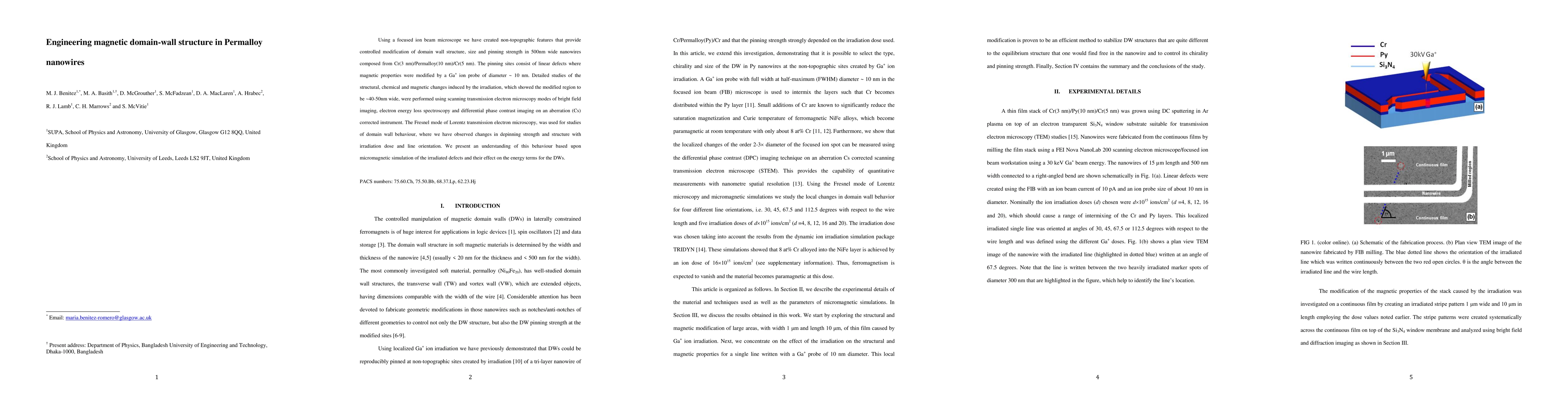
Using Lorentz transmission electron microscopy we investigate the behavior of domain walls pinned at non-topographic defects in Cr(3 nm)/Permalloy(10 nm)/Cr(5 nm) nanowires of width 500 nm. The pinning sites consist of linear defects where magnetic properties are modified by a Ga ion probe with diameter ~ 10 nm using a focused ion beam microscope. We study the detailed change of the modified region (which is on the scale of the focused ion spot) using electron energy loss spectroscopy and differential phase contrast imaging on an aberration (Cs) corrected scanning transmission electron microscope. The signal variation observed indicates that the region modified by the irradiation corresponds to ~ 40-50 nm despite the ion probe size of only 10 nm. Employing the Fresnel mode of Lorentz transmission electron microscopy, we show that it is possible to control the domain wall structure and its depinning strength not only via the irradiation dose but also the line orientation.
AI Key Findings
Get AI-generated insights about this paper's methodology, results, significance, and more — seven facets brought into focus.
Impact
Paper Details
PDF Preview
Citation Network
Current paper (gray), citations (green), references (blue)
Display is limited for performance on very large graphs.
Discussion 0