Enhancing Cardiac MRI Segmentation via Classifier-Guided Two-Stage Network and All-Slice Information Fusion Transformer
Publication
Metrics
AI Quick Summary
This paper proposes a classifier-guided two-stage network combined with an all-slice fusion transformer to improve cardiac MRI segmentation accuracy, especially for basal and apical slices. The method outperforms existing CNN-based and transformer-based models in Dice score and produces visually accurate segmentations.
Paper Preview
Abstract
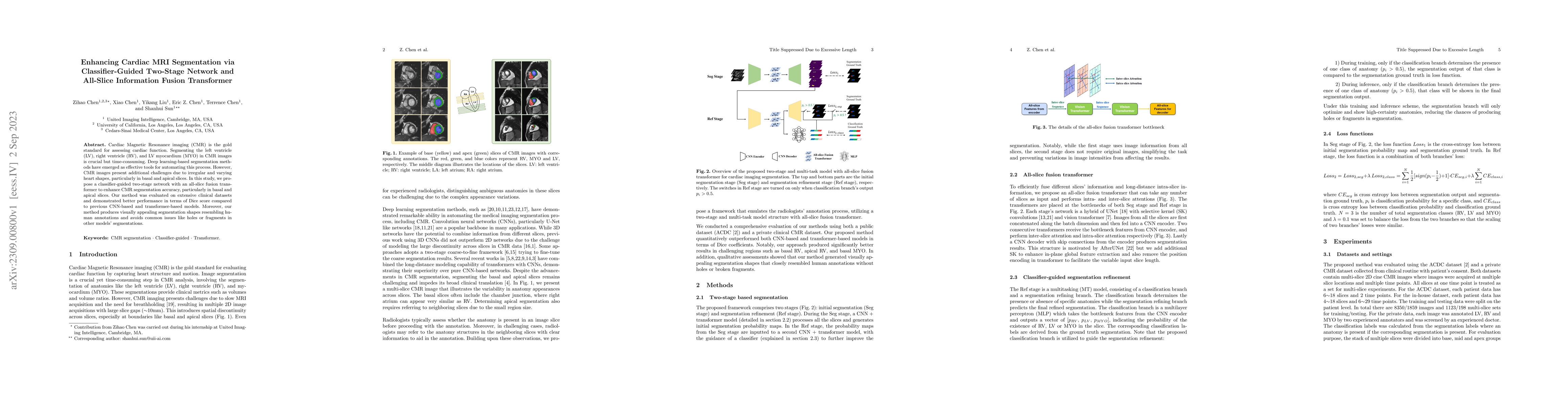
Cardiac Magnetic Resonance imaging (CMR) is the gold standard for assessing cardiac function. Segmenting the left ventricle (LV), right ventricle (RV), and LV myocardium (MYO) in CMR images is crucial but time-consuming. Deep learning-based segmentation methods have emerged as effective tools for automating this process. However, CMR images present additional challenges due to irregular and varying heart shapes, particularly in basal and apical slices. In this study, we propose a classifier-guided two-stage network with an all-slice fusion transformer to enhance CMR segmentation accuracy, particularly in basal and apical slices. Our method was evaluated on extensive clinical datasets and demonstrated better performance in terms of Dice score compared to previous CNN-based and transformer-based models. Moreover, our method produces visually appealing segmentation shapes resembling human annotations and avoids common issues like holes or fragments in other models' segmentations.
AI Key Findings
Get AI-generated insights about this paper's methodology, results, significance, and more — seven facets brought into focus.
Impact
Paper Details
Authors
PDF Preview
Key Terms
Citation Network
Current paper (gray), citations (green), references (blue)
Display is limited for performance on very large graphs.
Discussion 0