01
MethodologyHow they did it
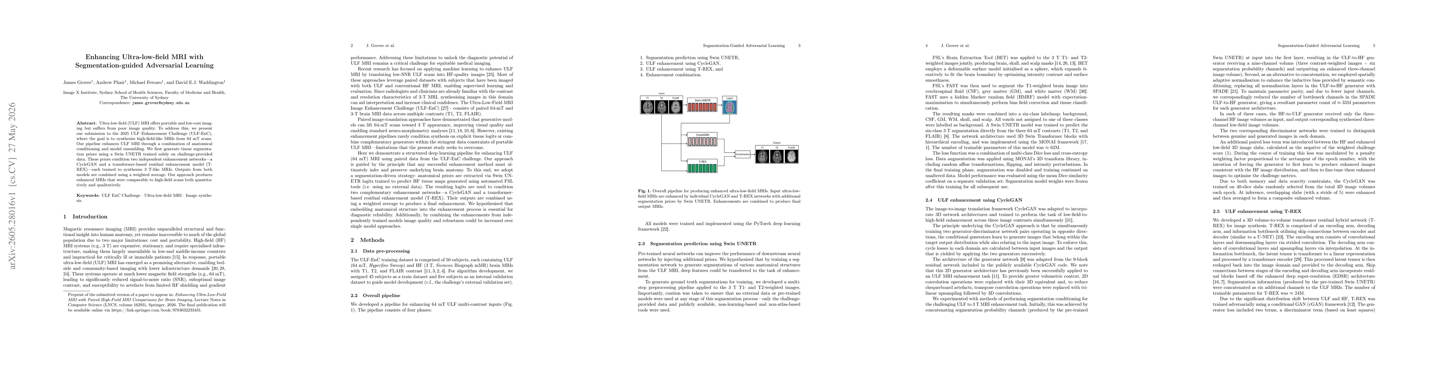
The study proposes a segmentation-guided enhancement pipeline for 64 mT ULFMRI. It first trains a SwinUNETR to generate tissue segmentation priors from challenge data, producing logits that act as anatomical conditioning. These priors condition two independent enhancement networks (CycleGAN and a transformer-based residual model, T-REX), each trained to synthesize 3T-like MRIs. Outputs from both models are combined via a weighted average to form the final enhanced image. The approach leverages paired ULFMRI–HF MRI data from the ULF-EnC dataset and emphasizes anatomical consistency to improve diagnostic reliability.
Discussion 0