Summary
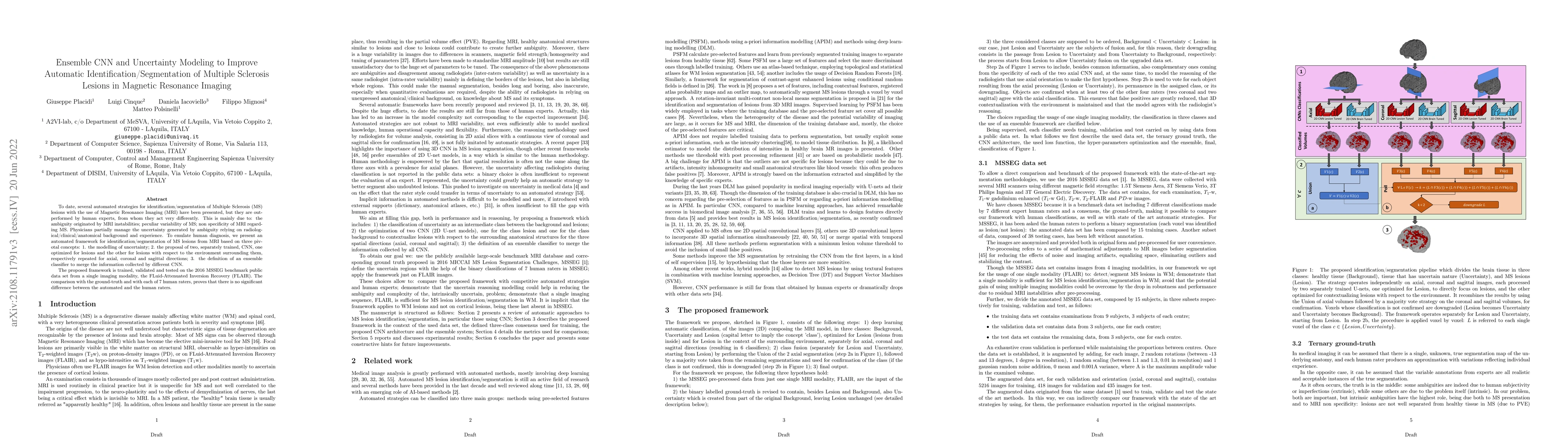
To date, several automated strategies for identification/segmentation of Multiple Sclerosis (MS) lesions with the use of Magnetic Resonance Imaging (MRI) have been presented, but they are outperformed by human experts, from whom they act very differently. This is mainly due to: the ambiguity originated by MRI instabilities; peculiar variability of MS; non specificity of MRI regarding MS. Physicians partially manage the uncertainty generated by ambiguity relying on radiological/clinical/anatomical background and experience. To emulate human diagnosis, we present an automated framework for identification/segmentation of MS lesions from MRI based on three pivotal concepts: 1. the modelling of uncertainty; 2. the proposal of two, separately trained, CNN, one optimized for lesions and the other for lesions with respect to the environment surrounding them, respectively repeated for axial, coronal and sagittal directions; 3. the definition of an ensemble classifier to merge the information collected by different CNN. The proposed framework is trained, validated and tested on the 2016 MSSEG benchmark public data set from a single imaging modality, the FLuid-Attenuated Inversion Recovery (FLAIR). The comparison with the ground-truth and with each of 7 human raters, proves that there is no significant difference between the automated and the human raters.
AI Key Findings
Get AI-generated insights about this paper's methodology, results, and significance.
Paper Details
PDF Preview
Key Terms
Citation Network
Current paper (gray), citations (green), references (blue)
Display is limited for performance on very large graphs.
Similar Papers
Found 4 papersSegHeD+: Segmentation of Heterogeneous Data for Multiple Sclerosis Lesions with Anatomical Constraints and Lesion-aware Augmentation
Wenjia Bai, Berke Doga Basaran, Paul M. Matthews
Interpretability of Uncertainty: Exploring Cortical Lesion Segmentation in Multiple Sclerosis
Henning Müller, Nataliia Molchanova, Alessandro Cagol et al.
| Title | Authors | Year | Actions |
|---|
Comments (0)