Ensemble of Pathology Foundation Models for MIDOG 2025 Track 2: Atypical Mitosis Classification
Publication
Metrics
Paper Preview
Abstract
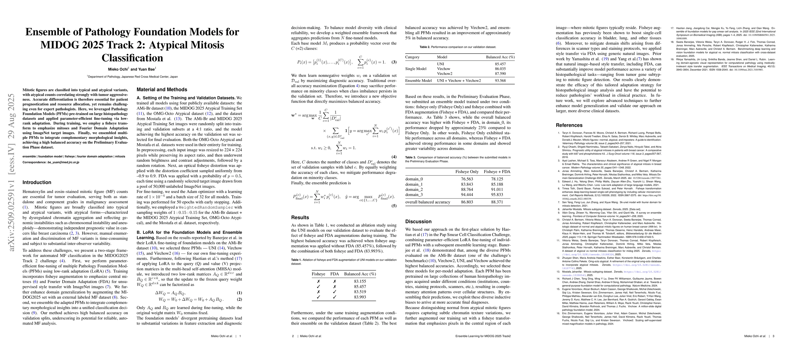
Mitotic figures are classified into typical and atypical variants, with atypical counts correlating strongly with tumor aggressiveness. Accurate differentiation is therefore essential for patient prognostication and resource allocation, yet remains challenging even for expert pathologists. Here, we leveraged Pathology Foundation Models (PFMs) pre-trained on large histopathology datasets and applied parameter-efficient fine-tuning via low-rank adaptation. In addition, we incorporated ConvNeXt V2, a state-of-the-art convolutional neural network architecture, to complement PFMs. During training, we employed a fisheye transform to emphasize mitoses and Fourier Domain Adaptation using ImageNet target images. Finally, we ensembled multiple PFMs to integrate complementary morphological insights, achieving competitive balanced accuracy on the Preliminary Evaluation Phase dataset.
AI Key Findings
Get AI-generated insights about this paper's methodology, results, significance, and more — seven facets brought into focus.
Discussion 0