Epicardial Adipose Tissue Segmentation from CT Images with A Semi-3D Neural Network
Publication
Metrics
AI Quick Summary
This paper proposes a U-Net-based semi-3D neural network for automatic segmentation of epicardial adipose tissue from CT scans, aiming to improve cardiovascular disease risk assessment. The method achieves a Dice score of 0.86 on 20 patient scans, demonstrating its potential for large-scale studies.
Paper Preview
Abstract
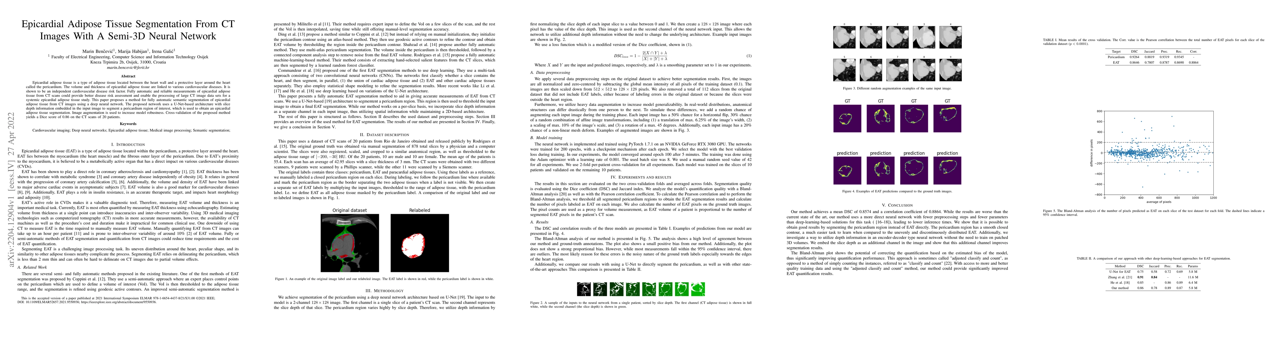
Epicardial adipose tissue is a type of adipose tissue located between the heart wall and a protective layer around the heart called the pericardium. The volume and thickness of epicardial adipose tissue are linked to various cardiovascular diseases. It is shown to be an independent cardiovascular disease risk factor. Fully automatic and reliable measurements of epicardial adipose tissue from CT scans could provide better disease risk assessment and enable the processing of large CT image data sets for a systemic epicardial adipose tissue study. This paper proposes a method for fully automatic semantic segmentation of epicardial adipose tissue from CT images using a deep neural network. The proposed network uses a U-Net-based architecture with slice depth information embedded in the input image to segment a pericardium region of interest, which is used to obtain an epicardial adipose tissue segmentation. Image augmentation is used to increase model robustness. Cross-validation of the proposed method yields a Dice score of 0.86 on the CT scans of 20 patients.
AI Key Findings
Get AI-generated insights about this paper's methodology, results, significance, and more — seven facets brought into focus.
Impact
Paper Details
Authors
PDF Preview
Key Terms
Citation Network
Current paper (gray), citations (green), references (blue)
Display is limited for performance on very large graphs.
Discussion 0