ArxivLens

Quick Summary:

This study proposes a machine learning method to estimate regional cerebral blood flow (CBF) using resting-state functional MRI (rsfMRI) data, demonstrating significant associations with actual CBF measured via arterial spin labeling (ASL). The method showed the highest correlation in the superior parietal lobule and correlated with cognitive scores, suggesting its potential as a surrogate for perfusion imaging when ASL is unavailable.

DOI: 10.1016/j.jneumeth.2019.108528
arXiv Id: 1907.08145
Comments: 20 pages, 6 main figures or tables, 4 supplementary figures or tables
Date Published: 2019-11-20
Date Updated: 2019-11-20

Key Terms (TF-IDF):

associations (0.248)
mri (0.243)
estimated (0.219)
score (0.218)
estimated (0.209)
score (0.207)
actual (0.205)
actual (0.204)
superior (0.193)
superior (0.191)
frequencies (0.186)
frequencies (0.183)
aging (0.156)
estimate (0.155)
machine (0.154)
estimate (0.150)
machine (0.150)
surrogate (0.145)
surrogate (0.144)
diseases (0.144)
License: http://arxiv.org/licenses/nonexclusive-distrib/1.0/

Summary:

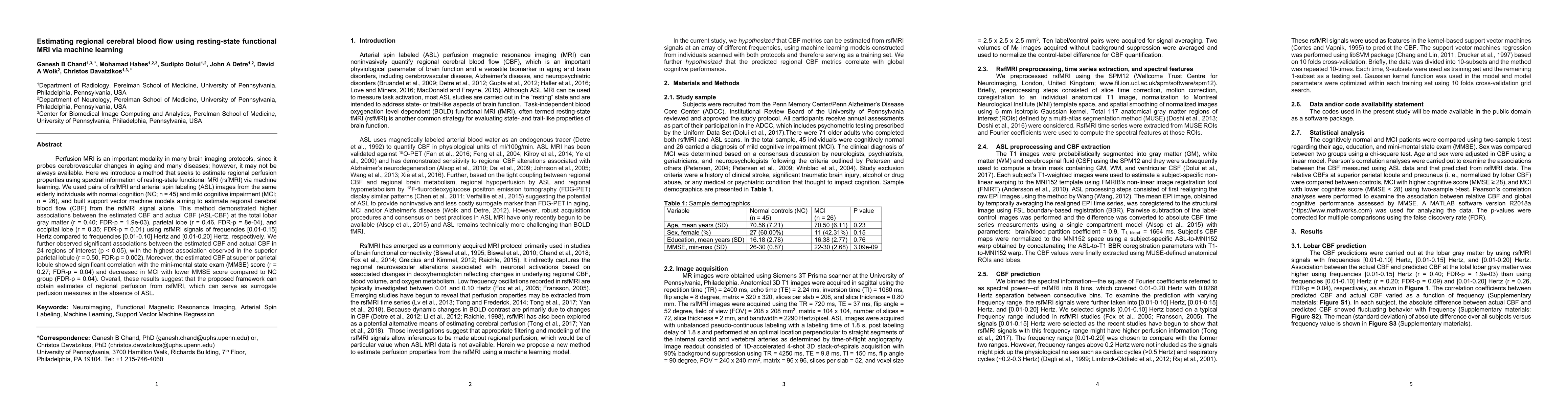
Perfusion MRI is an important modality in many brain imaging protocols, since it probes cerebrovascular changes in aging and many diseases; however, it may not be always available. Here we introduce a method that seeks to estimate regional perfusion properties using spectral information of resting-state functional MRI (rsfMRI) via machine learning. We used pairs of rsfMRI and arterial spin labeling (ASL) images from the same elderly individuals with normal cognition (NC; n = 45) and mild cognitive impairment (MCI; n = 26), and built support vector machine models aiming to estimate regional cerebral blood flow (CBF) from the rsfMRI signal alone. This method demonstrated higher associations between the estimated CBF and actual CBF (ASL-CBF) at the total lobar gray matter (r = 0.40; FDR-p = 1.9e-03), parietal lobe (r = 0.46, FDR-p = 8e-04), and occipital lobe (r = 0.35; FDR-p = 0.01) using rsfMRI signals of frequencies [0.01-0.15] Hertz compared to frequencies [0.01-0.10] Hertz and [0.01-0.20] Hertz, respectively. We further observed significant associations between the estimated CBF and actual CBF in 24 regions of interest (p < 0.05), with the highest association observed in the superior parietal lobule (r = 0.50, FDR-p = 0.002). Moreover, the estimated CBF at superior parietal lobule showed significant correlation with the mini-mental state exam (MMSE) score (r = 0.27; FDR-p = 0.04) and decreased in MCI with lower MMSE score compared to NC group (FDR-p = 0.04). Overall, these results suggest that the proposed framework can obtain estimates of regional perfusion from rsfMRI, which can serve as surrogate perfusion measures in the absence of ASL.

Citation & Reference Network Visualization

This graph shows how this paper is connected to other research. The current paper is shown in dark gray, papers that cite it are in green, and papers it references are in blue. You can:

Current Paper
Papers citing this work (Citations)
Papers cited by this work (References)

• Hover over any node to see the paper title

• Click a node to view that paper's details

• Drag nodes to rearrange the graph

• Use the zoom controls (bottom right) to zoom in/out

Citations and References

Title Authors Year Actions
AgeNet-SHAP: An explainable AI approach for optimally mapping multivariate regional brain age and clinical severity patterns in Alzheimer’s disease
Gauri Darekar, Taslim Murad, Hui-Yuan Miao, Deepa S. Thakuri, Ganesh B. Chand
2025
View Paper
Characterizing multivariate regional hubs for schizophrenia classification, sex differences, and brain age estimation using explainable AI
Yuzheng Nie, Taslim Murad, Hui-Yuan Miao, Puskar Bhattarai, Deepa S. Thakuri, Ganesh B. Chand
2025
View Paper
Effect of carotid artery stenting on cognitive function in patients with asymptomatic carotid artery stenosis, a multimodal magnetic resonance study.
Dangzhen Wang, Juan Xia, Liang Li, Tao Wang
2024
View Paper
Explainable AI-based Deep-SHAP for mapping the multivariate relationships between regional neuroimaging biomarkers and cognition.
Puskar Bhattarai, Deepa S. Thakuri, Yuzheng Nie, Ganesh B. Chand
2024
View Paper
Predicting cognitive dysfunction and regional hubs using Braak staging amyloid-beta biomarkers and machine learning
Puskar Bhattarai, Ahmed Taha, Bhavin Soni, Deepa S. Thakuri, Erin Ritter, Ganesh B. Chand
2023
View Paper
Salience network anatomical and molecular markers are linked with cognitive dysfunction in mild cognitive impairment
G. Chand, Deepa S. Thakuri, Bhavin Soni
2022
View Paper
Brain Tumor detection Using Machine Learning and Deep Learning Approaches
Oneza Tehreem Khan, D. Rajeswari
2022
View Paper
Neuro-anatomical and neuro-molecular markers in salience network and their integration in machine learning-based prediction of cognitive dysfunction in mild cognitive impairment
G. Chand, Deepa S. Thakuri, Bhavin Soni
2021
View Paper
Stress Changes the Resting-State Cortical Flow of Information from Distributed to Frontally Directed Patterns
Soheil Keshmiri
2020
View Paper
Becker Digital
Puskar Bhattarai, Ahmed Taha, Bhavin Soni, Deepa S. Thakuri, Erin Ritter, Ganesh B. Chand
View Paper

Similar Papers

PINNing Cerebral Blood Flow: Analysis of Perfusion MRI in Infants using Physics-Informed Neural Networks

Ching-En Chiu, Marta Varela, Christoforos Galazis, Anil A. Bharath, Tomoki Arichi

Published: 2024-10-11

Machine learning for cerebral blood vessels' malformations

Jean Barbier, Édgar Roldán, Roman Belousov, Deniz Eroglu, Irem Topal, Alexander Cherevko, Yuri Bugay, Maxim Shishlenin

Published: 2024-11-25

Fast Cerebral Blood Flow Analysis via Extreme Learning Machine

Xi Chen, Zhenya Zang, Xingda Li

Published: 2024-02-02

Comments (0)

This website uses cookies to ensure you get the best experience. Learn more