Publication
Metrics
AI Quick Summary
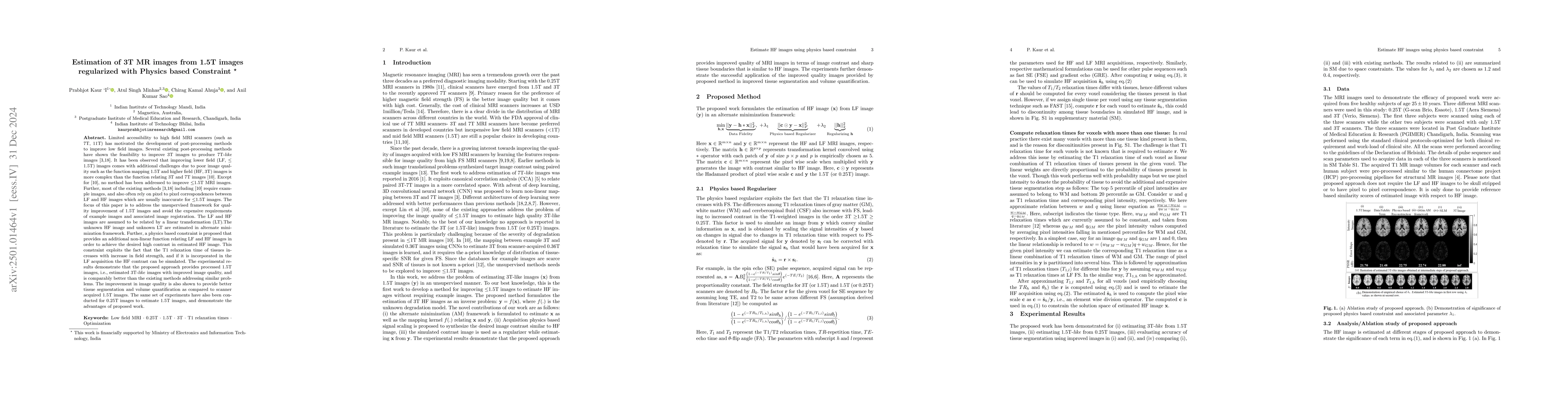
This paper proposes an unsupervised method to enhance 1.5T MRI images to produce 3T-like images without requiring example images or image registration. It uses a linear transformation and a physics-based constraint to estimate high-contrast, improved 3T-like images, demonstrating better tissue segmentation and volume quantification compared to original 1.5T images.
Paper Preview
Abstract
Limited accessibility to high field MRI scanners (such as 7T, 11T) has motivated the development of post-processing methods to improve low field images. Several existing post-processing methods have shown the feasibility to improve 3T images to produce 7T-like images [3,18]. It has been observed that improving lower field (LF, <=1.5T) images comes with additional challenges due to poor image quality such as the function mapping 1.5T and higher field (HF, 3T) images is more complex than the function relating 3T and 7T images [10]. Except for [10], no method has been addressed to improve <=1.5T MRI images. Further, most of the existing methods [3,18] including [10] require example images, and also often rely on pixel to pixel correspondences between LF and HF images which are usually inaccurate for <=1.5T images. The focus of this paper is to address the unsupervised framework for quality improvement of 1.5T images and avoid the expensive requirements of example images and associated image registration. The LF and HF images are assumed to be related by a linear transformation (LT). The unknown HF image and unknown LT are estimated in alternate minimization framework. Further, a physics based constraint is proposed that provides an additional non-linear function relating LF and HF images in order to achieve the desired high contrast in estimated HF image. The experimental results demonstrate that the proposed approach provides processed 1.5T images, i.e., estimated 3T-like images with improved image quality, and is comparably better than the existing methods addressing similar problems. The improvement in image quality is also shown to provide better tissue segmentation and volume quantification as compared to scanner acquired 1.5T images.
AI Key Findings
Get AI-generated insights about this paper's methodology, results, significance, and more — seven facets brought into focus.
Paper Details
Authors
PDF Preview
Related Papers
No references found for this paper.
Discussion 0