Quick Summary:

This study uses high-resolution 4DCT imaging to acquire dynamic contrast enhancement maps of the left atrial appendage (LAA) during multiple heartbeats, enabling estimation of blood flow parameters through computational modeling. The results provide insights into spatial-temporal flow characteristics, which are crucial for evaluating patients with Atrial Fibrillation.

Summary

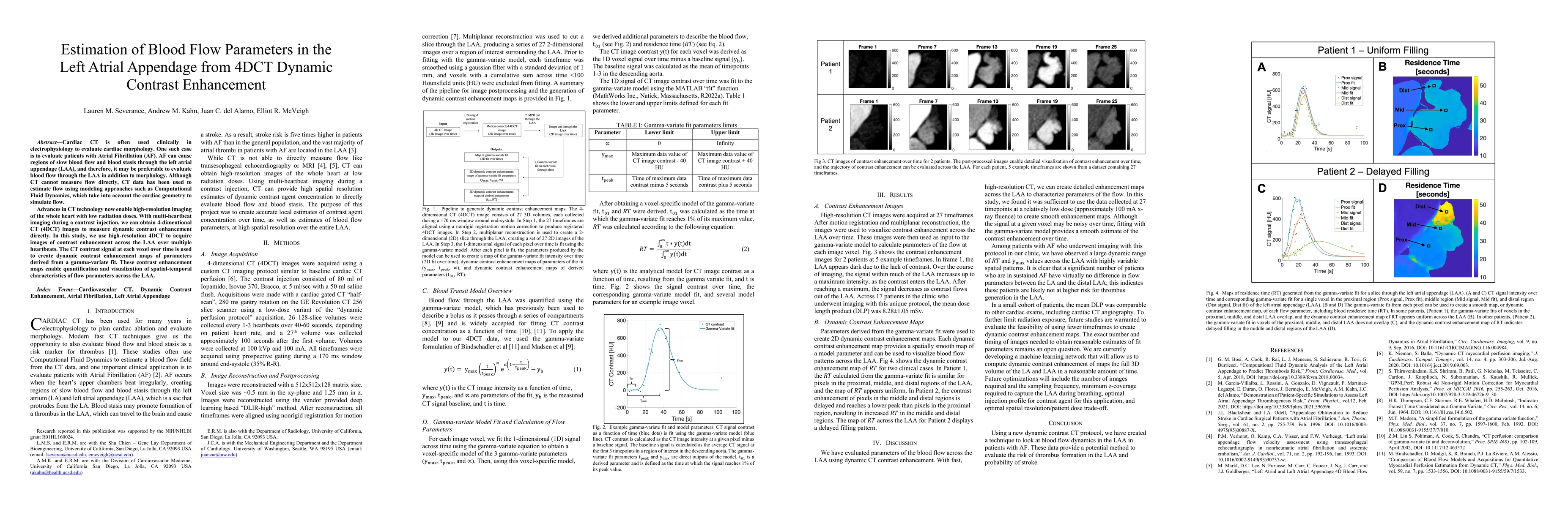
Cardiac CT is often used clinically in electrophysiology to evaluate cardiac morphology. One such case is to evaluate patients with Atrial Fibrillation (AF). AF can cause regions of slow blood flow and blood stasis through the left atrial appendage (LAA), and therefore, it may be preferable to evaluate blood flow through the LAA in addition to morphology. Although CT cannot measure flow directly, CT data has been used to estimate flow using modeling approaches such as Computational Fluid Dynamics, which take into account the cardiac geometry to simulate flow. Advances in CT technology now enable high-resolution imaging of the whole heart with low radiation doses. With multi-heartbeat imaging during a contrast injection, we can obtain 4-dimentional CT (4DCT) images to measure dynamic contrast enhancement directly. In this study, we use high-resolution 4DCT to acquire images of contrast enhancement across the LAA over multiple heartbeats. The CT contrast signal at each voxel over time is used to create dynamic contrast enhancement maps of parameters derived from a gamma-variate fit. These contrast enhancement maps enable quantification and visualization of spatial-temporal characteristics of flow parameters across the LAA.

AI Key Findings

Generated Jun 11, 2025

Methodology

The study employed high-resolution 4DCT to capture contrast enhancement across the Left Atrial Appendage (LAA) over multiple heartbeats. The CT contrast signal at each voxel over time was utilized to generate dynamic contrast enhancement maps of parameters derived from a gamma-variate fit, enabling quantification and visualization of flow parameters across the LAA.

Key Results

  • High-resolution CT images were acquired at 27 timeframes, providing detailed visualization of contrast enhancement over time across the LAA.
  • Dynamic contrast enhancement maps were created using parameters derived from the gamma-variate fit, allowing for visualization of blood flow patterns across the LAA.
  • A large dynamic range of residence time (RT) and y!"# values was observed across the LAA in patients with atrial fibrillation, indicating variability in thrombus risk.
  • Mean DLP was comparable to other cardiac exams, including cardiac CT angiography, suggesting the feasibility of using this protocol for thrombus risk evaluation.
  • A machine learning network is being developed to compute dynamic contrast enhancement maps of the full 3D volume of the LA and LAA efficiently.

Significance

This research offers a novel method to evaluate the risk of thrombus formation in the LAA and predict stroke probability in patients with atrial fibrillation using dynamic contrast CT protocols.

Technical Contribution

The study presents a new dynamic contrast CT protocol and a gamma-variate model for quantifying blood flow parameters in the LAA, providing high-resolution, spatially smooth enhancement maps.

Novelty

This work introduces a novel application of 4DCT and gamma-variate modeling to assess blood flow dynamics in the LAA, offering a potential method for evaluating thrombus risk in atrial fibrillation patients.

Limitations

  • The study did not compare the proposed method directly with other established techniques for flow evaluation.
  • The optimal number of timeframes and sampling frequency for reasonable estimates of fit parameters remains to be determined.

Future Work

  • Investigate the feasibility of using fewer timeframes to create dynamic contrast enhancement maps while maintaining accuracy.
  • Optimize the number of images required and sampling frequency, as well as the minimum z-coverage needed to capture the LAA during breathing.
  • Determine the optimal injection profile for contrast agent and spatial resolution/patient dose trade-off.

Paper Details

PDF Preview

Download PDF

Key Terms

No key terms available for this paper.

No citations found for this paper.

Comments (0)

This website uses cookies to ensure you get the best experience. Learn more