Evaluation of Deep Learning-based Scatter Correction for Total-body PET
Publication
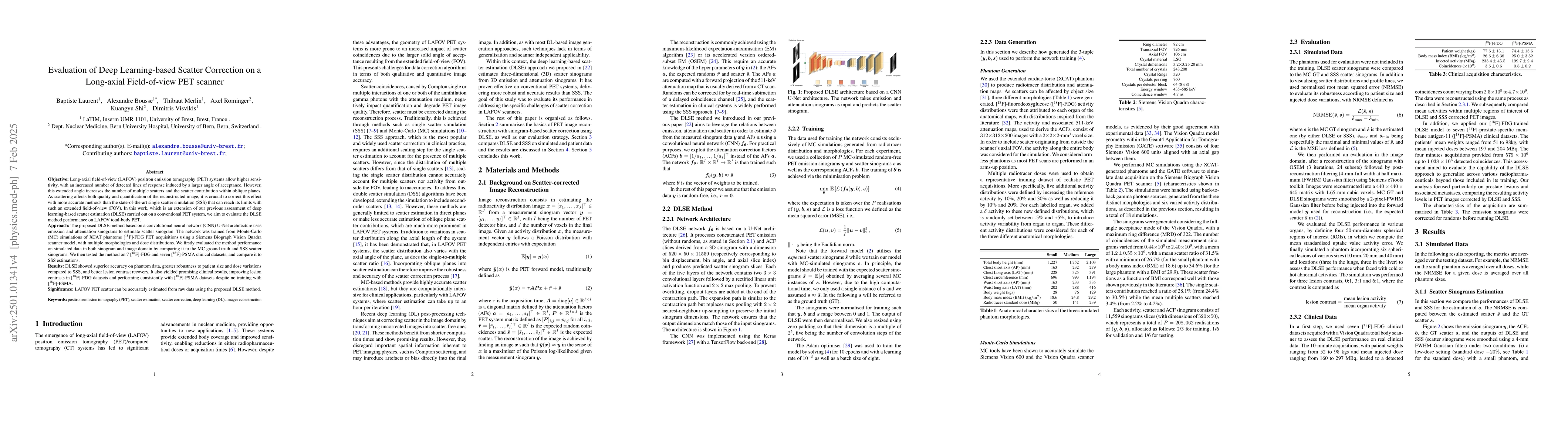
Metrics
AI Quick Summary
This paper evaluates a deep learning-based scatter correction method (DLSE) using a CNN U-Net architecture for long-axial field-of-view total-body PET, showing superior accuracy and robustness compared to single scatter simulation (SSS), and improving lesion contrast in clinical [18F]-FDG datasets.
Paper Preview
Abstract
Objective: Long-axial field-of-view (LAFOV) positron emission tomography (PET) systems allow higher sensitivity, with an increased number of detected lines of response induced by a larger angle of acceptance. However this extend angle increase the number of multiple scatters and the scatter contribution within oblique planes. As scattering affects both quality and quantification of the reconstructed image, it is crucial to correct this effect with more accurate methods than the state-of-the-art single scatter simulation (SSS) that can reach its limits with such an extended field-of-view (FOV). In this work, which is an extension of our previous assessment of deep learning-based scatter estimation (DLSE) carried out on a conventional PET system, we aim to evaluate the DLSE method performance on LAFOV total-body PET. Approach: The proposed DLSE method based on an convolutional neural network (CNN) U-Net architecture uses emission and attenuation sinograms to estimate scatter sinogram. The network was trained from Monte-Carlo (MC) simulations of XCAT phantoms [18F]-FDG PET acquisitions using a Siemens Biograph Vision Quadra scanner model, with multiple morphologies and dose distributions. We firstly evaluated the method performance on simulated data in both sinogram and image domain by comparing it to the MC ground truth and SSS scatter sinograms. We then tested the method on 7 [18F]-FDG and 7 [18F]-PSMA clinical datasets, and compare it to SSS estimations. Results: DLSE showed superior accuracy on phantom data, greater robustness to patient size and dose variations compared to SSS, and better lesion contrast recovery. It also yielded promising clinical results, improving lesion contrasts in [18 F]-FDG datasets and performing consistently with [18F]-PSMA datasets despite no training with [18F]-PSMA.
AI Key Findings
Get AI-generated insights about this paper's methodology, results, significance, and more — seven facets brought into focus.
Impact
Paper Details
Authors
PDF Preview
Citation Network
Current paper (gray), citations (green), references (blue)
Display is limited for performance on very large graphs.
Discussion 0