EXACT-CT: EXplainable Analysis for Crohn's and Tuberculosis using CT

Source: ArXiv

AI Quick Summary

This research develops EXACT-CT, a method using 3D CT scans, computer vision, and machine learning to differentiate between Crohn's disease and intestinal tuberculosis, which often present overlapping features. The study employs XGBoost with SHAP analysis to identify key radiological biomarkers and compares its effectiveness against state-of-the-art methods.

Share: Share on X LinkedIn

Summary

Crohn's disease and intestinal tuberculosis share many overlapping features such as clinical, radiological, endoscopic, and histological features - particularly granulomas, making it challenging to clinically differentiate them. Our research leverages 3D CTE scans, computer vision, and machine learning to improve this differentiation to avoid harmful treatment mismanagement such as unnecessary anti-tuberculosis therapy for Crohn's disease or exacerbation of tuberculosis with immunosuppressants. Our study proposes a novel method to identify radiologist - identified biomarkers such as VF to SF ratio, necrosis, calcifications, comb sign and pulmonary TB to enhance accuracy. We demonstrate the effectiveness by using different ML techniques on the features extracted from these biomarkers, computing SHAP on XGBoost for understanding feature importance towards predictions, and comparing against SOTA methods such as pretrained ResNet and CTFoundation.
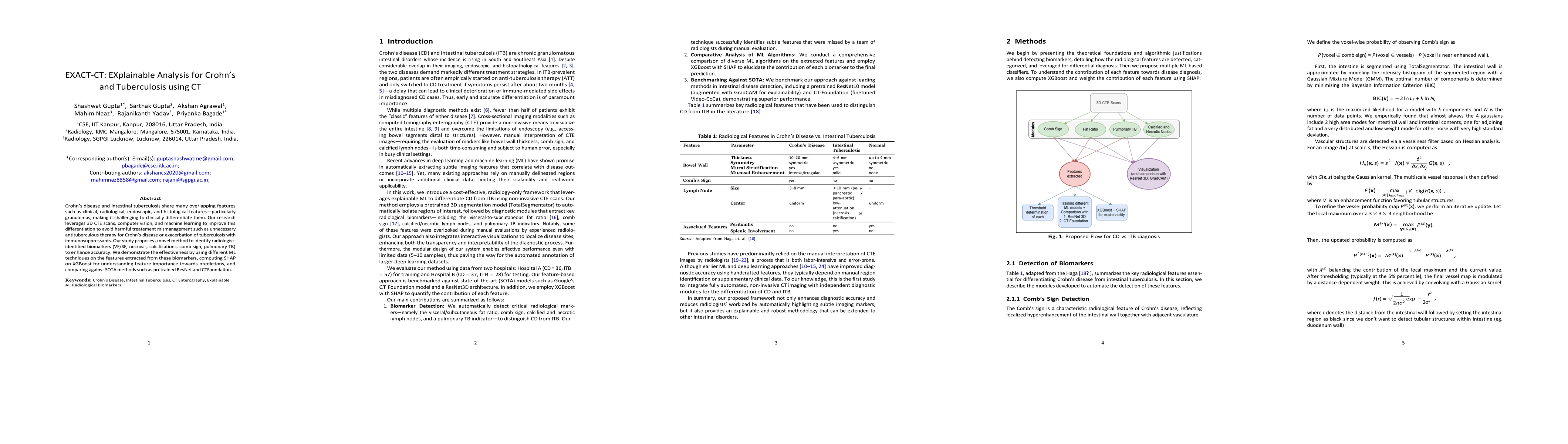
AI Key Findings

Generated Jun 10, 2025

Methodology

The research proposes a novel method, EXACT-CT, that combines classical image processing techniques with deep learning methods to automatically extract and analyze key radiological biomarkers for differentiating Crohn's Disease (CD) from Intestinal Tuberculosis (ITB) using CT Enterography images.

Key Results

  • EXACT-CT achieves competitive diagnostic performance compared to state-of-the-art 3D deep learning models like ResNet3D and CTFoundation.
  • The framework effectively identifies biomarkers such as visceral-to-subcutaneous fat ratio, Comb's sign, and evidence of pulmonary tuberculosis, as well as proxy detectors for necrotic and calcified lymph nodes.
  • The accompanying 3D visualization tool aids radiologists by highlighting subtle imaging features that can be missed in high-volume workflows.
  • Hand-crafted features capturing core radiological biomarkers can achieve competitive performance, underscoring the continued value of interpretable features in clinical decision-making.

Significance

This research can reduce diagnostic ambiguity, minimize reliance on empirical treatment trials, and ultimately improve patient outcomes by providing an explainable framework for differentiating CD from ITB using CT Enterography images.

Technical Contribution

The development of EXACT-CT, a novel and explainable framework for differentiating CD from ITB using CT Enterography images, combining classical image processing techniques with deep learning methods.

Novelty

EXACT-CT offers competitive performance with state-of-the-art deep learning models while providing clear visual and numerical insights for clinical decision-making, bridging the gap between classical computer vision and advanced AI methods.

Limitations

  • The dataset is relatively small and sourced from only two South Asian hospitals, which may limit the generalizability of findings.
  • Some modules rely on empirically set parameters that might not be optimal under varying imaging conditions or across diverse patient populations.
  • Proxy detectors for necrotic and calcified lymph nodes produce a significant number of false positives and need further refinement.

Future Work

  • Optimize implementation for parallel processing and dynamic hyperparameter tuning methods.
  • Incorporate additional radiological biomarkers and validate algorithms on diverse datasets from geographically varied populations.
  • Integrate deep learning approaches that automatically learn optimal features and explore hybrid models combining classical image processing, deep learning, and multimodal data.
  • Collaborate with HCI experts to design intuitive interfaces and conduct clinical trials for real-world validation.

Paper Details

PDF Preview

Download PDF

Citation Network

Current paper (gray), citations (green), references (blue)

Display is limited for performance on very large graphs.

Current Paper
Citations
References
Click to view

No citations found for this paper.

Comments (0)

This website uses cookies to ensure you get the best experience. Learn more