Publication
Metrics
Paper Preview
Abstract
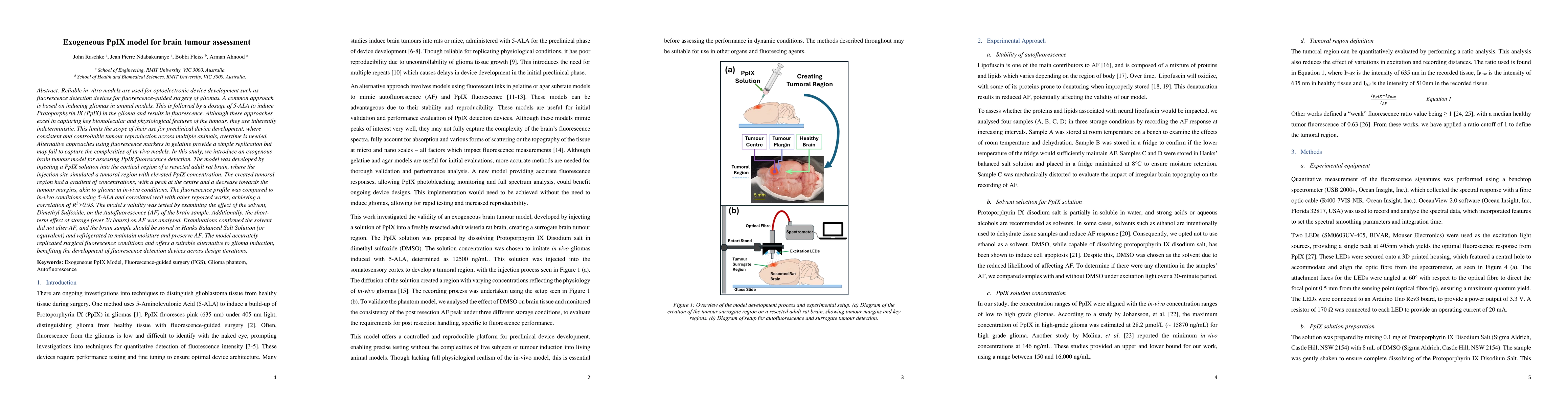
Reliable in-vitro models are used for optoelectronic device development such as fluorescence detection devices for fluorescence-guided surgery of gliomas. A common approach is based on inducing gliomas in animal models. This is followed by a dosage of 5-ALA to induce Protoporphyrin IX (PpIX) in the glioma and which fluoresces. Although these approaches excel in capturing key biomolecular and physiological features of the tumour, they are inherently indeterministic. This limits the scope of their use for preclinical device development, where consistent and controllable tumour reproduction across multiple animals is needed. Approaches using fluorescence markers in gelatine provide a simple replication but fail to capture the complexities of in-vivo models. In this study, we introduce an exogenous brain tumour model for assessing PpIX fluorescence detection. The model was developed by injecting a PpIX solution into the cortical region of a resected adult rat brain, the injection site simulated a tumoral region with elevated PpIX concentration. The tumoral region had a gradient of concentrations, with a peak at the centre and a decrease towards the margins, akin to in-vivo gliomas. The fluorescence profile was compared to in-vivo conditions using 5-ALA and correlated well with other reported works, achieving a correlation of R2>0.93. The model's validity was tested by examining the effect of the solvent, DMSO, on the Autofluorescence (AF) of the brain sample and the short-term effect of storage on AF was analysed. Examinations confirmed the solvent did not alter AF, and the brain sample should be stored in Hanks Balanced Salt Solution and refrigerated to maintain moisture and preserve AF. The model accurately replicated surgical fluorescence conditions and offers a suitable alternative to glioma induction, benefiting the development of fluorescence detection devices across design iterations.
AI Key Findings
Get AI-generated insights about this paper's methodology, results, significance, and more — seven facets brought into focus.
Discussion 0