This paper demonstrates experimental 3D coherent diffractive imaging using photon-sparse random projections from a synchrotron X-ray source, providing a valuable analog for XFEL imaging. It shows that a sparsity of $1.3\times10^{-3}$ photons per pixel can be overcome, offering crucial insights for atomic-resolution single particle imaging with XFELs.
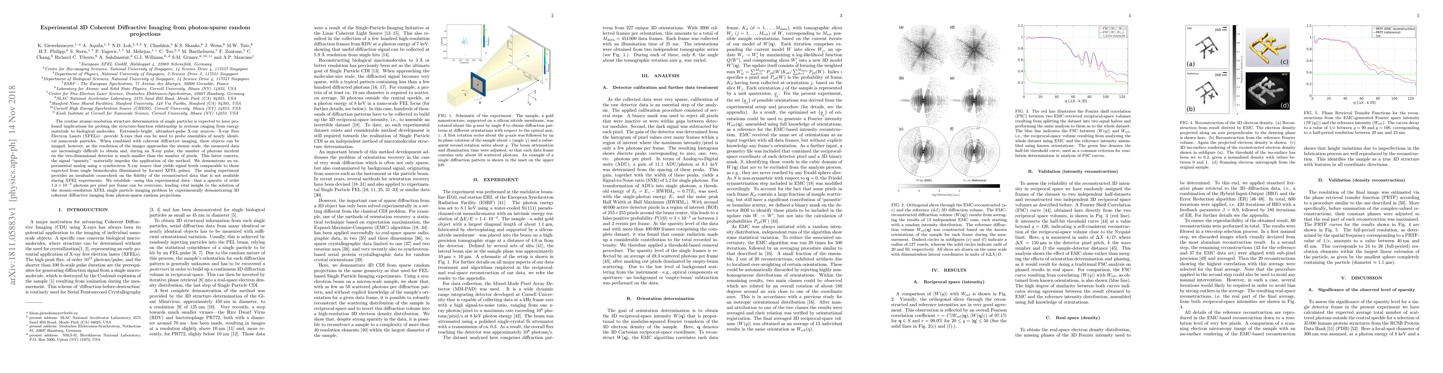
The routine atomic-resolution structure determination of single particles is expected to have profound implications for probing the structure-function relationship in systems ranging from energy materials to biological molecules. Extremely-bright, ultrashort-pulse X-ray sources---X-ray Free Electron Lasers (XFELs)---provide X-rays that can be used to probe ensembles of nearly identical nano-scale particles. When combined with coherent diffractive imaging, these objects can be imaged; however, as the resolution of the images approaches the atomic scale, the measured data are increasingly difficult to obtain and, during an X-ray pulse, the number of photons incident on the two-dimensional detector is much smaller than the number of pixels. This latter concern, the signal "sparsity," materially impedes the application of the method. We demonstrate an experimental analog using a synchrotron X-ray source that yields signal levels comparable to those expected from single biomolecules illuminated by focused XFEL pulses. The analog experiment provides an invaluable cross-check on the fidelity of the reconstructed data that is not available during XFEL experiments. We establish---using this experimental data---that a sparsity of order $1.3\times10^{-3}$ photons per pixel per frame can be overcome, lending vital insight to the solution of the atomic-resolution XFEL single particle imaging problem by experimentally demonstrating 3D coherent diffractive imaging from photon-sparse random projections.
This graph shows how this paper is connected to other research. The current paper is shown in dark gray, papers that cite it are in green, and papers it references are in blue. You can:
• Hover over any node to see the paper title
• Click a node to view that paper's details
• Drag nodes to rearrange the graph
• Use the zoom controls (bottom right) to zoom in/out
Title | Authors | Year | Actions |
---|---|---|---|
Molecular size dependence on achievable resolution from XFEL single-particle 3D reconstruction
|
M. Nakano, O. Miyashita, F. Tama
|
2023
|
View Paper |
Expected resolution limits of x-ray free-electron laser single-particle imaging for realistic source and detector properties
|
J. E, Y. Kim, J. Bielecki, M. Sikorski, R. de Wijn, C. Fortmann-Grote, J. Sztuk-Dambietz, J. Koliyadu, R. Letrun, H. Kirkwood, T. Sato, R. Bean, A. Mancuso, C. Kim
|
2022
|
View Paper |
Characterization of 128 × 128 MM-PAD-2.1 ASIC: a fast framing hard x-ray detector with high dynamic range
|
D. Gadkari, K. Shanks, H. Hu, H. Philipp, M. Tate, J. Thom-Levy, S. Gruner
|
2022
|
View Paper |
Single-image super-resolution improvement of X-ray single-particle diffraction images using convolutional neural network
|
Atsushi Tokuhisa, Yoshinobu Akinaga, Kei Terayama, Yasushi Okuno
|
2021
|
View Paper |
Characterization of 128x128 MM-PAD-2.1 ASIC: A Fast Framing Hard X-Ray Detector with High Dynamic Range
|
D. Gadkari, K. Shanks, H. Hu, H. Philipp, M. Tate, J. Thom-Levy, S. Gruner
|
2021
|
View Paper |
Determination of the Structure of Biological Macromolecular Particles Using X-Ray Lasers. Achievements and Prospects
|
T. Petrova, V. Lunin
|
2020
|
View Paper |
An encryption–decryption framework to validating single-particle imaging
|
Zhou Shen, C. Teo, K. Ayyer, N. Loh
|
2020
|
View Paper |
3D diffractive imaging of nanoparticle ensembles using an x-ray laser
|
K. Ayyer, P. L. Xavier, J. Bielecki, Zhou Shen, B. Daurer, A. Samanta, S. Awel, Richard Bean, A. Barty, T. Ekeberg, Armando D. Estillore, K. Giewekemeyer, M. Hunter, R. Kirian, H. Kirkwood, Yoonhee Kim, Jayanath C. P. Koliyadu, H. Lange, Romain Letruin, J. Lubke, A. Morgan, N. Roth, Tokushi Sato, M. Sikorski, F. Schulz, J. Spence, P. Vagovič, T. Wollweber, L. Worbs, O. Yefanov, Yu-De Zhuang, F. Maia, D. Horke, J. Kupper, N. Loh, A. Mancuso, H. Chapman
|
2020
|
View Paper |
Single-shot transverse coherence measurements with Young's double pinholes at FLASH2
|
T. Wodzinski, M. Mehrjoo, M. Ruiz-Lopez, B. Keitel, M. Kuhlmann, M. Brachmanski, S. Künzel, M. Fajardo, E. Plönjes
|
2020
|
View Paper |
Perspectives on single particle imaging with x rays at the advent of high repetition rate x-ray free electron laser sources
|
J. Bielecki, F. Maia, A. Mancuso
|
2020
|
View Paper |
High dynamic range CdTe mixed-mode pixel array detector (MM-PAD) for kilohertz imaging of hard x-rays
|
H. Philipp, M. Tate, K. Shanks, P. Purohit, S. Gruner
|
2020
|
View Paper |
Reference-enhanced x-ray single-particle imaging
|
K. Ayyer
|
2020
|
View Paper |
X-Ray Tomography
|
C. Jacobsen
|
2019
|
View Paper |
X-Ray Microscope Systems
|
C. Jacobsen
|
2019
|
View Paper |
X-Ray Focusing Optics
|
C. Jacobsen
|
2019
|
View Paper |
X-Ray Physics
|
C. Jacobsen
|
2019
|
View Paper |
X-Ray Microscope Instrumentation
|
C. Jacobsen
|
2019
|
View Paper |
X-ray Microscopy
|
C. Jacobsen
|
2019
|
View Paper |
Characterization of a Fast-Framing X-Ray Camera With Wide Dynamic Range for High-Energy Imaging
|
K. Shanks, H. Philipp, J. Weizeorick, M. Hammer, M. Tate, P. Purohit, A. Miceli, S. Gruner
|
2019
|
View Paper |
Reconstruction of 3D structure for nanoscale biological objects from experiments data on super-bright X-ray free electron lasers (XFELs): dependence of the 3D resolution on the experiment parameters
|
K. Ikonnikova, A. Teslyuk, S. Bobkov, S. Zolotarev, V. Ilyin
|
2019
|
View Paper |
3D diffractive imaging of nanoparticle ensembles using an x-ray laser
|
N. And
|
View Paper |
Comments (0)