Exploration of Multi-Scale Image Fusion Systems in Intelligent Medical Image Analysis
Publication
Metrics
AI Quick Summary
This research develops a U-Net-based MRI segmentation algorithm that incorporates residual networks and enhanced context information to improve brain tumor segmentation accuracy. Experimental results show an impressive 3D reconstruction accuracy of 0.9851, significantly enhancing both segmentation precision and classification efficiency.
Paper Preview
Abstract
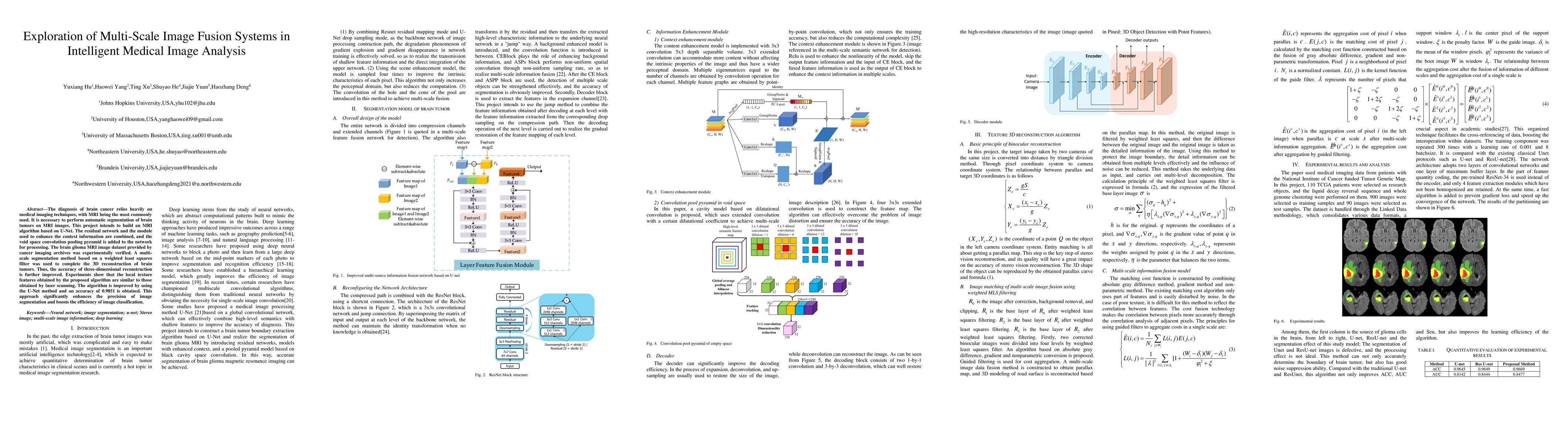
The diagnosis of brain cancer relies heavily on medical imaging techniques, with MRI being the most commonly used. It is necessary to perform automatic segmentation of brain tumors on MRI images. This project intends to build an MRI algorithm based on U-Net. The residual network and the module used to enhance the context information are combined, and the void space convolution pooling pyramid is added to the network for processing. The brain glioma MRI image dataset provided by cancer imaging archives was experimentally verified. A multi-scale segmentation method based on a weighted least squares filter was used to complete the 3D reconstruction of brain tumors. Thus, the accuracy of three-dimensional reconstruction is further improved. Experiments show that the local texture features obtained by the proposed algorithm are similar to those obtained by laser scanning. The algorithm is improved by using the U-Net method and an accuracy of 0.9851 is obtained. This approach significantly enhances the precision of image segmentation and boosts the efficiency of image classification.
AI Key Findings
Get AI-generated insights about this paper's methodology, results, significance, and more — seven facets brought into focus.
Impact
Paper Details
Authors
PDF Preview
Key Terms
Citation Network
Current paper (gray), citations (green), references (blue)
Display is limited for performance on very large graphs.
Discussion 0