Exploring Regions of Interest: Visualizing Histological Image Classification for Breast Cancer using Deep Learning
Publication
Metrics
AI Quick Summary
This research investigates the use of convolutional neural networks (CNNs) to classify breast cancer in histological images, focusing on visualizing regions of interest to improve the interpretability of deep learning models. The study employs the VGG19 architecture and various visualization and pixel selection methods to compare CNN-identified regions with those marked by pathologists. The Gradient visualization and MeanShift selection methods proved most effective in this endeavor.
Paper Preview
Abstract
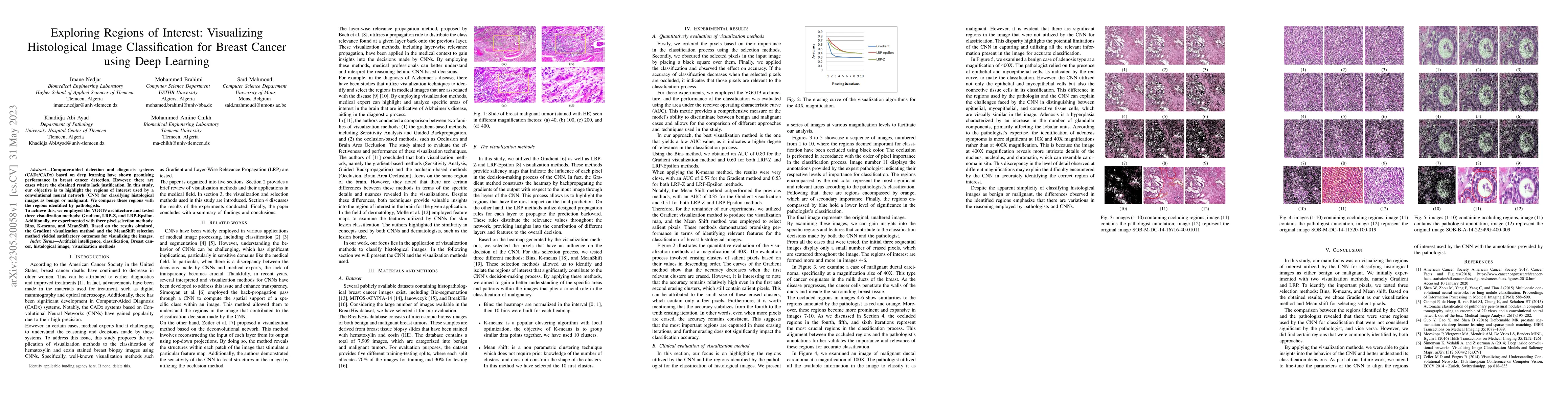
Computer aided detection and diagnosis systems based on deep learning have shown promising performance in breast cancer detection. However, there are cases where the obtained results lack justification. In this study, our objective is to highlight the regions of interest used by a convolutional neural network (CNN) for classifying histological images as benign or malignant. We compare these regions with the regions identified by pathologists. To achieve this, we employed the VGG19 architecture and tested three visualization methods: Gradient, LRP Z, and LRP Epsilon. Additionally, we experimented with three pixel selection methods: Bins, K-means, and MeanShift. Based on the results obtained, the Gradient visualization method and the MeanShift selection method yielded satisfactory outcomes for visualizing the images.
AI Key Findings
Get AI-generated insights about this paper's methodology, results, significance, and more — seven facets brought into focus.
Impact
Paper Details
Authors
PDF Preview
Key Terms
Citation Network
Current paper (gray), citations (green), references (blue)
Display is limited for performance on very large graphs.
Discussion 0