01
MethodologyHow they did it
The research uses a U-Net architecture for lesion segmentation from whole-body FDG-PET/CT scans.
This paper investigates the application of a vanilla U-Net for segmenting tumor lesions in whole-body FDG-PET/CT scans, focusing on network architecture, data preprocessing, and data augmentation. The study demonstrates that the vanilla U-Net can achieve competitive results, securing first place in the autoPET 2022 challenge leaderboards.
This paper investigates the application of a vanilla U-Net for segmenting tumor lesions in whole-body FDG-PET/CT scans, focusing on network architecture, data preprocessing, and data augmentation. The study demonstrates that the vanilla U-Net can achieve competitive results, securing first place in the autoPET 2022 challenge leaderboards.
The research uses a U-Net architecture for lesion segmentation from whole-body FDG-PET/CT scans. More in Methodology →
Achieves first place in both preliminary and final leaderboards of the autoPET 2022 challenge — Vanilla U-Net with proper input shape achieves satisfactory performance More in Key Results →
This research is important because it contributes to better tumor segmentation results using FDG-PET/CT scans. More in Significance →
Data augmentation may not be sufficient to improve performance — Inference time may vary depending on input shape and step size More in Limitations →
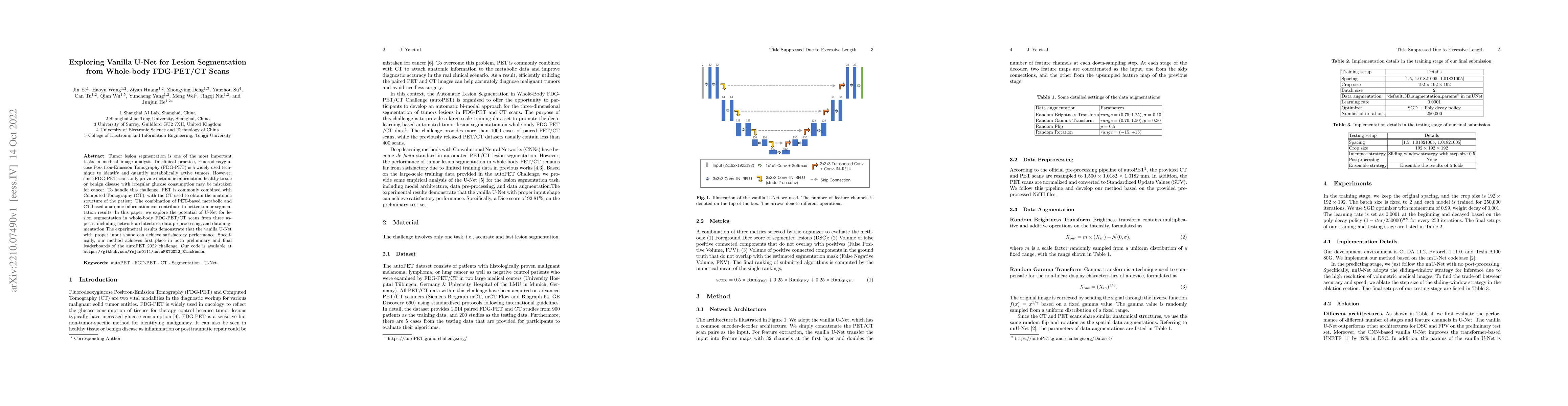
Tumor lesion segmentation is one of the most important tasks in medical image analysis. In clinical practice, Fluorodeoxyglucose Positron-Emission Tomography~(FDG-PET) is a widely used technique to identify and quantify metabolically active tumors. However, since FDG-PET scans only provide metabolic information, healthy tissue or benign disease with irregular glucose consumption may be mistaken for cancer. To handle this challenge, PET is commonly combined with Computed Tomography~(CT), with the CT used to obtain the anatomic structure of the patient. The combination of PET-based metabolic and CT-based anatomic information can contribute to better tumor segmentation results. %Computed tomography~(CT) is a popular modality to illustrate the anatomic structure of the patient. The combination of PET and CT is promising to handle this challenge by utilizing metabolic and anatomic information. In this paper, we explore the potential of U-Net for lesion segmentation in whole-body FDG-PET/CT scans from three aspects, including network architecture, data preprocessing, and data augmentation. The experimental results demonstrate that the vanilla U-Net with proper input shape can achieve satisfactory performance. Specifically, our method achieves first place in both preliminary and final leaderboards of the autoPET 2022 challenge. Our code is available at https://github.com/Yejin0111/autoPET2022_Blackbean.
Seven facets of this paper, analysed and brought into focus by AI.
This research is important because it contributes to better tumor segmentation results using FDG-PET/CT scans.
The research uses a U-Net architecture for lesion segmentation from whole-body FDG-PET/CT scans.
This research is important because it contributes to better tumor segmentation results using FDG-PET/CT scans.
The research presents a novel approach to lesion segmentation using U-Net architecture.
This work is different from existing research because it uses a U-Net architecture specifically designed for whole-body FDG-PET/CT scans
Current paper (gray), citations (green), references (blue)
Display is limited for performance on very large graphs.
Discussion 0