Publication
Metrics
AI Quick Summary
This paper explores extending the depth of field in high-resolution scanning transmission electron microscopy (STEM) to image nanoparticles in focus. It demonstrates the application of optical microscopy techniques to achieve a 6 nm depth of field in a 100 kV aberration-corrected STEM, enabling clear imaging of Pt-Co nanoparticles on a carbon support.
Paper Preview
Abstract
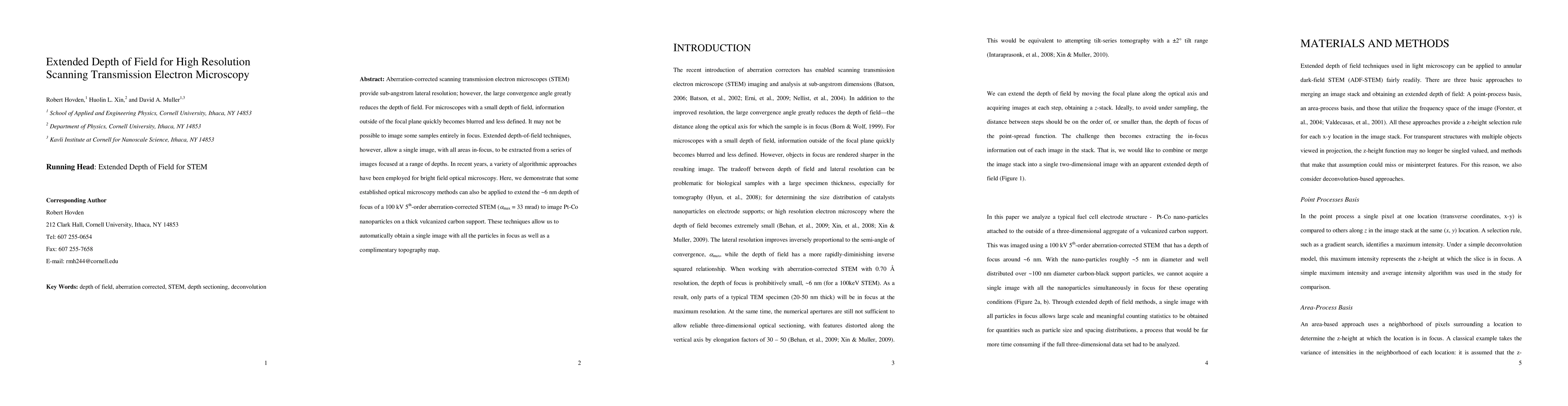
Aberration-corrected scanning transmission electron microscopes (STEM) provide sub-angstrom lateral resolution; however, the large convergence angle greatly reduces the depth of field. For microscopes with a small depth of field, information outside of the focal plane quickly becomes blurred and less defined. It may not be possible to image some samples entirely in focus. Extended depth-of-field techniques, however, allow a single image, with all areas in-focus, to be extracted from a series of images focused at a range of depths. In recent years, a variety of algorithmic approaches have been employed for bright field optical microscopy. Here, we demonstrate that some established optical microscopy methods can also be applied to extend the ~6 nm depth of focus of a 100 kV 5th-order aberration-corrected STEM (alpha_max = 33 mrad) to image Pt-Co nanoparticles on a thick vulcanized carbon support. These techniques allow us to automatically obtain a single image with all the particles in focus as well as a complimentary topography map.
AI Key Findings
Get AI-generated insights about this paper's methodology, results, significance, and more — seven facets brought into focus.
Impact
Paper Details
PDF Preview
Key Terms
Citation Network
Current paper (gray), citations (green), references (blue)
Display is limited for performance on very large graphs.
Discussion 0