Summary
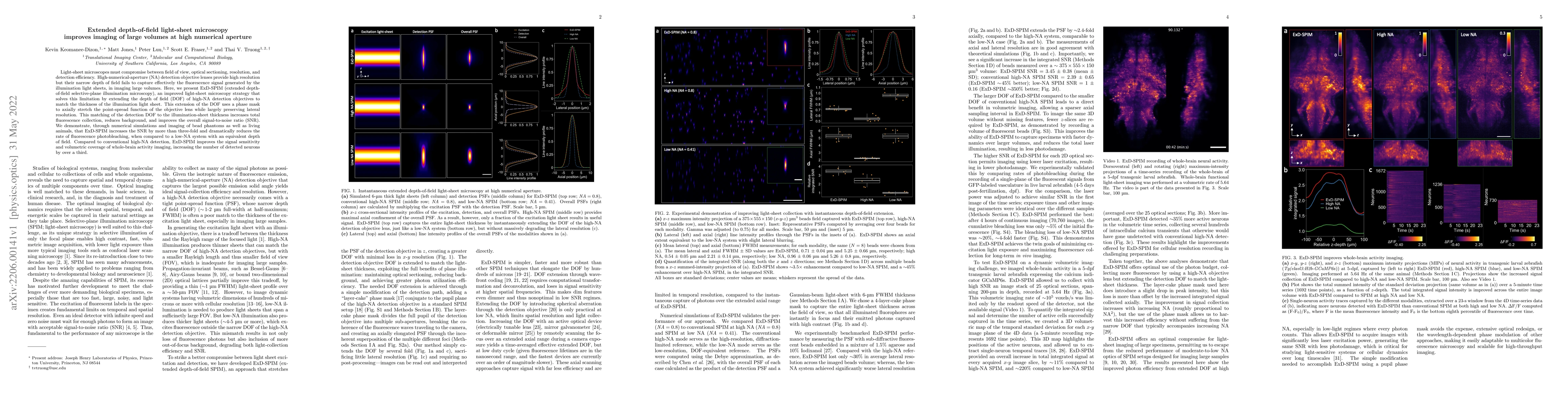
Light-sheet microscopes must compromise between field of view, optical sectioning, resolution, and detection efficiency. High-numerical-aperture (NA) detection objective lenses provide high resolution but their narrow depth of field fails to capture effectively the fluorescence signal generated by the illumination light sheets, in imaging large volumes. Here, we present ExD-SPIM (extended depth-of-field selective-plane illumination microscopy), an improved light-sheet microscopy strategy that solves this limitation by extending the depth of field (DOF) of high-NA detection objectives to match the thickness of the illumination light sheet. This extension of the DOF uses a phase mask to axially stretch the point-spread function of the objective lens while largely preserving lateral resolution. This matching of the detection DOF to the illumination-sheet thickness increases total fluorescence collection, reduces background, and improves the overall signal-to-noise ratio (SNR). We demonstrate, through numerical simulations and imaging of bead phantoms as well as living animals, that ExD-SPIM increases the SNR by more than three-fold and dramatically reduces the rate of fluorescence photobleaching, when compared to a low-NA system with an equivalent depth of field. Compared to conventional high-NA detection, ExD-SPIM improves the signal sensitivity and volumetric coverage of whole-brain activity imaging, increasing the number of detected neurons by over a third.
AI Key Findings
Get AI-generated insights about this paper's methodology, results, and significance.
Paper Details
PDF Preview
Key Terms
Citation Network
Current paper (gray), citations (green), references (blue)
Display is limited for performance on very large graphs.
Similar Papers
Found 4 papersSlanted light-sheet array microscopy for large volume imaging at rates exceeding 100 Hz
Junyi Li, Nanguang Chen, Anqi Qiu et al.
Full-aperture extended-depth oblique plane microscopy through dynamic remote focusing
Paolo Pozzi, Vipin Balan, Alessia Candeo et al.
| Title | Authors | Year | Actions |
|---|
Comments (0)