Extracting, Visualizing, and Learning from Dynamic Data: Perfusion in Surgical Video for Tissue Characterization
Publication
Metrics
AI Quick Summary
This paper presents a method for characterizing cancerous versus benign rectal lesions using early fluorescence imaging from indocyanine green (ICG) administration during surgery. The approach involves signal processing and visualization techniques to extract and analyze dynamic data from surgical videos for improved tissue characterization.
Paper Preview
Abstract
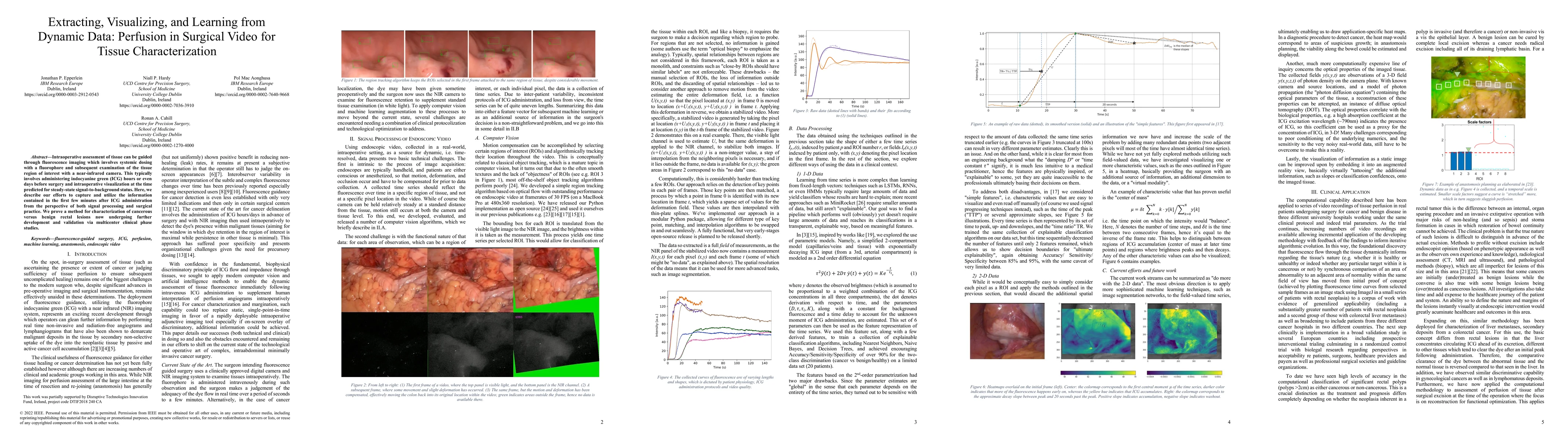
Intraoperative assessment of tissue can be guided through fluorescence imaging which involves systemic dosing with a fluorophore and subsequent examination of the tissue region of interest with a near-infrared camera. This typically involves administering indocyanine green (ICG) hours or even days before surgery and intraoperative visualization at the time predicted for steady-state signal-to-background status. Here, we describe our efforts to capture and utilize the information contained in the first few minutes after ICG administration from the perspective of both signal processing and surgical practice. We prove a method for characterization of cancerous versus benign rectal lesions now undergoing further development and validation via multicenter clinical phase studies.
AI Key Findings
Get AI-generated insights about this paper's methodology, results, significance, and more — seven facets brought into focus.
Impact
Paper Details
Authors
PDF Preview
Key Terms
Citation Network
Current paper (gray), citations (green), references (blue)
Display is limited for performance on very large graphs.
Discussion 0