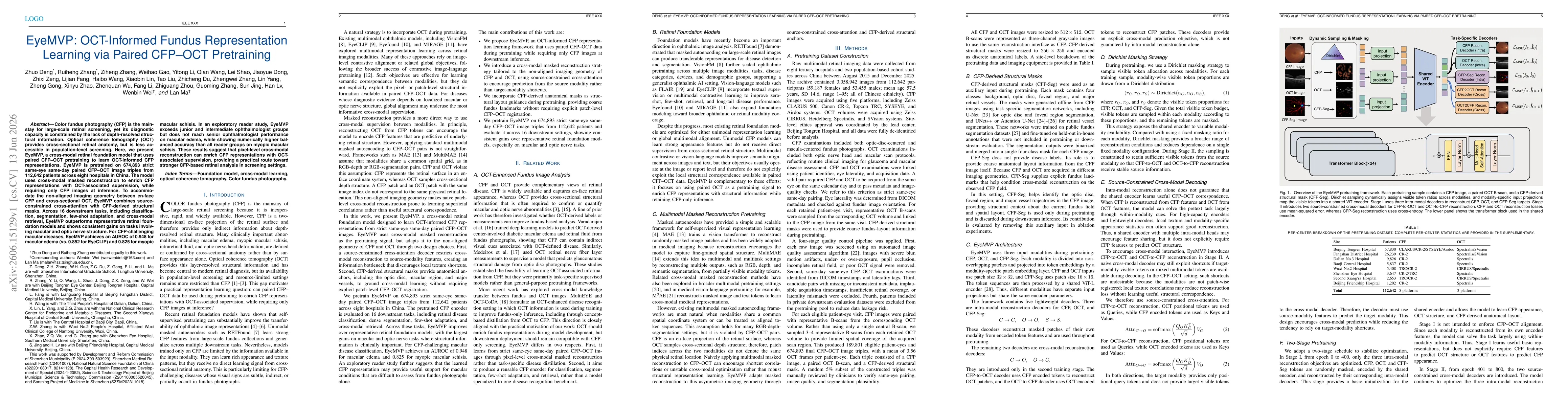
EyeMVP is a cross-modal retinal foundation model pretrained on 674,893 strict same-eye, same-day paired CFP–OCT image triples from 112,642 patients across eight hospitals in China. It uses cross-modal masked reconstruction to enrich CFP representations with OCT supervision while inference uses only CFP inputs. To handle non-aligned CFP and OCT geometry, it employs (i) source-constrained cross-attention decoders to restrict cross-modal reconstruction to source modality features and (ii) CFP-derived anatomical masks (optic disc, macular region, major vessels) as structural guidance. The model is trained with a two-stage schedule and includes a CFP-Seg branch to provide coarse anatomical guidance without requiring patch-level CFP–OCT registration. Evaluation spans 16 downstream tasks including classification, segmentation, few-shot adaptation, and cross-modal retrieval, demonstrating consistent improvements over baseline retinal foundation models, particularly in macular and optic nerve structure-related tasks.
EyeMVP: OCT-Informed Fundus Representation Learning via Paired CFP--OCT Pretraining
Publication
Metrics
Quick Answers
What methodology did the authors use?
EyeMVP is a cross-modal retinal foundation model pretrained on 674,893 strict same-eye, same-day paired CFP–OCT image triples from 112,642 patients across eight hospitals in China. It uses cross-modal masked reconstruction to enrich CFP representations with OCT supervision while inference uses only CFP inputs. To handle non-aligned CFP and OCT geometry, it employs (i) source-constrained cross-attention decoders to restrict cross-modal reconstruction... More in Methodology →
What are the key results?
EyeMVP achieves higher performance than representative retinal foundation models across 16 downstream tasks, with largest gains on macular and optic nerve structure–related tasks. — For CFP-based macular disease classification, EyeMVP attains AUROC of 0.948 for macular edema and 0.825 for myopic macular schisis, outperforming EyeCLIP (0.852). More in Key Results →
Why is this work significant?
Demonstrates that pixel-level cross-modal reconstruction leveraging paired CFP–OCT data can enrich CFP representations with OCT-associated supervision, enabling stronger CFP-based retinal analysis in screening settings without needing OCT at deployment; highlights potential for improved detection and characterization of macular and optic nerve pathology in large-scale screening. More in Significance →
What are the main limitations?
Pretraining data are from Chinese cohorts; external validation across diverse populations, devices, and healthcare settings is needed. — Baseline comparisons are not data-matched in pretraining scale and disease distribution, so some gains may reflect data advantages rather than architectural effects. More in Limitations →
Paper Preview
Abstract
Color fundus photography (CFP) is the mainstay for large-scale retinal screening, yet its diagnostic capacity is constrained by the lack of depth-resolved structural information. Optical coherence tomography (OCT) provides cross-sectional retinal anatomy, but is less accessible in population-level screening. Here, we present EyeMVP, a cross-modal retinal foundation model that uses paired CFP--OCT pretraining to learn OCT-informed CFP representations. EyeMVP is pretrained on 674,893 strict same-eye same-day paired CFP--OCT image triples from 112,642 patients across eight hospitals in China. The model uses cross-modal masked reconstruction to enrich CFP representations with OCT-associated supervision, while requiring only CFP images at inference. To accommodate the non-aligned imaging geometry between en-face CFP and cross-sectional OCT, EyeMVP combines source-constrained cross-attention with CFP-derived structural masks. Across 16 downstream tasks, including classification, segmentation, few-shot adaptation, and cross-modal retrieval, EyeMVP outperforms representative retinal foundation models and shows consistent gains on tasks involving macular and optic nerve structure. For CFP-challenging macular diseases, EyeMVP achieves an AUROC of 0.948 for macular edema (vs.~0.852 for EyeCLIP) and 0.825 for myopic macular schisis. In an exploratory reader study, EyeMVP exceeds junior and intermediate ophthalmologist groups but does not reach senior ophthalmologist performance on macular edema, while showing numerically higher balanced accuracy than all reader groups on myopic macular schisis. These results suggest that pixel-level cross-modal reconstruction can enrich CFP representations with OCT-associated supervision, providing a practical route toward stronger CFP-based retinal analysis in screening settings.
Key Findings, in focus
Seven facets of this paper, analysed and brought into focus by AI.
Demonstrates that pixel-level cross-modal reconstruction leveraging paired CFP–OCT data can enrich CFP representations with OCT-associated supervision, enabling stronger CFP-based retinal analysis in screening settings without needing OCT at deployment; highlights potential for improved detection and characterization of macular and optic nerve pathology in large-scale screening.
- EyeMVP achieves higher performance than representative retinal foundation models across 16 downstream tasks, with largest gains on macular and optic nerve structure–related tasks.
- For CFP-based macular disease classification, EyeMVP attains AUROC of 0.948 for macular edema and 0.825 for myopic macular schisis, outperforming EyeCLIP (0.852).
- In an exploratory reader study, EyeMVP surpasses junior and intermediate ophthalmologists for macular edema and shows numerically higher balanced accuracy than all reader groups for myopic macular schisis.
- The approach demonstrates effective OCT-informed CFP representation learning via pixel-level cross-modal reconstruction without requiring OCT at inference.
Demonstrates that pixel-level cross-modal reconstruction leveraging paired CFP–OCT data can enrich CFP representations with OCT-associated supervision, enabling stronger CFP-based retinal analysis in screening settings without needing OCT at deployment; highlights potential for improved detection and characterization of macular and optic nerve pathology in large-scale screening.
Introduction of EyeMVP, an OCT-informed CFP representation learning framework that (a) uses strict same-eye, same-day paired CFP–OCT pretraining, (b) applies source-constrained cross-modal decoding to handle non-aligned CFP and OCT geometry, and (c) incorporates CFP-derived anatomical masks to ground cross-modal learning, resulting in a reusable CFP encoder for classification, segmentation, few-shot learning, and cross-modal retrieval.
First to learn cross-modal OCT-informed CFP representations from strict same-eye paired CFP–OCT data with a non-aligned geometry, using source-constrained cross-attention and CFP-derived structural masks to enable effective pixel-level cross-modal supervision for broad downstream retinal tasks.
- Pretraining data are from Chinese cohorts; external validation across diverse populations, devices, and healthcare settings is needed.
- Baseline comparisons are not data-matched in pretraining scale and disease distribution, so some gains may reflect data advantages rather than architectural effects.
- Current model uses static CFP and OCT images; longitudinal modeling of disease progression is not explored.
- eye-level linking lacks exact spatial CFP–OCT correspondence for precise regional inference.
- Prospective external validation across institutions and device types, with calibrated uncertainty estimation.
- Incorporation of longitudinal data to model disease progression and temporal dynamics.
- Workflow integration studies for CFP-based screening/triage incorporating uncertainty estimates.
- Exploration of extending source-constrained cross-attention ideas to other non-aligned multimodal medical pairs.
Discussion 0