Fast and Automatic Periacetabular Osteotomy Fragment Pose Estimation Using Intraoperatively Implanted Fiducials and Single-View Fluoroscopy
Publication
Metrics
Paper Preview
Abstract
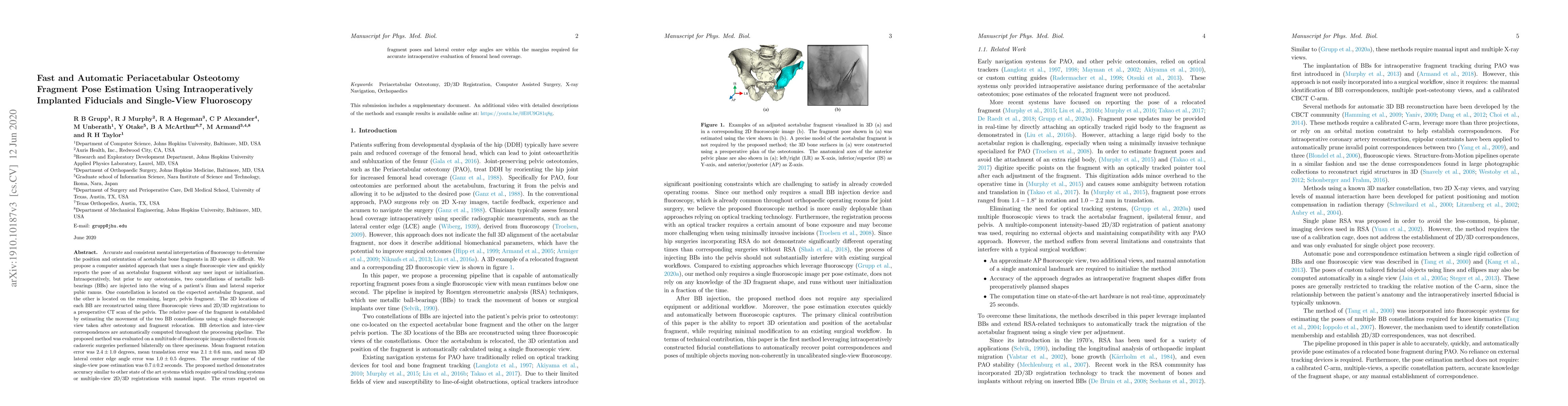
Accurate and consistent mental interpretation of fluoroscopy to determine the position and orientation of acetabular bone fragments in 3D space is difficult. We propose a computer assisted approach that uses a single fluoroscopic view and quickly reports the pose of an acetabular fragment without any user input or initialization. Intraoperatively, but prior to any osteotomies, two constellations of metallic ball-bearings (BBs) are injected into the wing of a patient's ilium and lateral superior pubic ramus. One constellation is located on the expected acetabular fragment, and the other is located on the remaining, larger, pelvis fragment. The 3D locations of each BB are reconstructed using three fluoroscopic views and 2D/3D registrations to a preoperative CT scan of the pelvis. The relative pose of the fragment is established by estimating the movement of the two BB constellations using a single fluoroscopic view taken after osteotomy and fragment relocation. BB detection and inter-view correspondences are automatically computed throughout the processing pipeline. The proposed method was evaluated on a multitude of fluoroscopic images collected from six cadaveric surgeries performed bilaterally on three specimens. Mean fragment rotation error was 2.4 +/- 1.0 degrees, mean translation error was 2.1 +/- 0.6 mm, and mean 3D lateral center edge angle error was 1.0 +/- 0.5 degrees. The average runtime of the single-view pose estimation was 0.7 +/- 0.2 seconds. The proposed method demonstrates accuracy similar to other state of the art systems which require optical tracking systems or multiple-view 2D/3D registrations with manual input. The errors reported on fragment poses and lateral center edge angles are within the margins required for accurate intraoperative evaluation of femoral head coverage.
AI Key Findings
Get AI-generated insights about this paper's methodology, results, significance, and more — seven facets brought into focus.
Impact
Paper Details
PDF Preview
Key Terms
Citation Network
Current paper (gray), citations (green), references (blue)
Display is limited for performance on very large graphs.
Discussion 0