Fast and label-free 3D virtual H&E histology via active modulation-assisted dynamic full-field OCT
Publication
Metrics
AI Quick Summary
This research introduces active phase modulation-assisted dynamic full-field OCT (APMD-FFOCT) to enhance imaging stability and contrast in fresh tissue, enabling conversion to virtual H&E-stained images via unsupervised deep learning. The method facilitates rapid, 3D virtual histology at a rate of 1 frame per second, proving its potential in intraoperative cancer diagnosis for central nervous system and breast tissues.
Paper Preview
Abstract
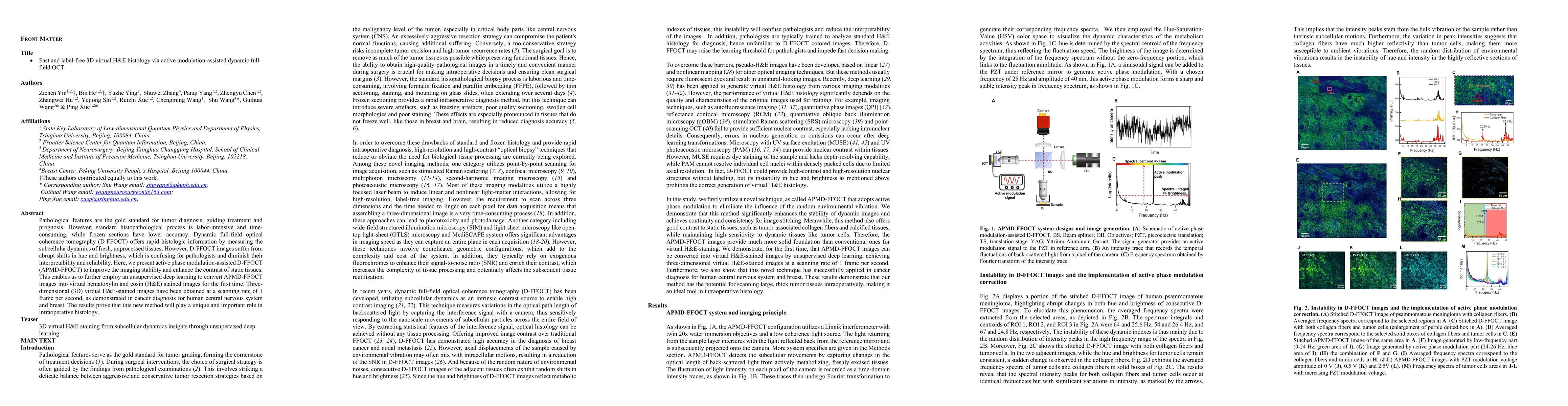
Pathological features are the gold standard for tumor diagnosis, guiding treatment and prognosis. However, standard histopathological process is labor-intensive and time-consuming, while frozen sections have lower accuracy. Dynamic full-field optical coherence tomography (D-FFOCT) offers rapid histologic information by measuring the subcellular dynamics of fresh, unprocessed tissues. However, D-FFOCT images suffer from abrupt shifts in hue and brightness, which is confusing for pathologists and diminish their interpretability and reliability. Here, we present active phase modulation-assisted D-FFOCT (APMD-FFOCT) to improve the imaging stability and enhance the contrast of static tissues. This enables us to further employ an unsupervised deep learning to convert APMD-FFOCT images into virtual hematoxylin and eosin (H&E) stained images for the first time. Three-dimensional (3D) virtual H&E-stained images have been obtained at a scanning rate of 1 frame per second, as demonstrated in cancer diagnosis for human central nervous system and breast. The results prove that this new method will play a unique and important role in intraoperative histology.
AI Key Findings
Get AI-generated insights about this paper's methodology, results, significance, and more — seven facets brought into focus.
Impact
Paper Details
Authors
PDF Preview
Key Terms
Citation Network
Current paper (gray), citations (green), references (blue)
Display is limited for performance on very large graphs.
Discussion 0