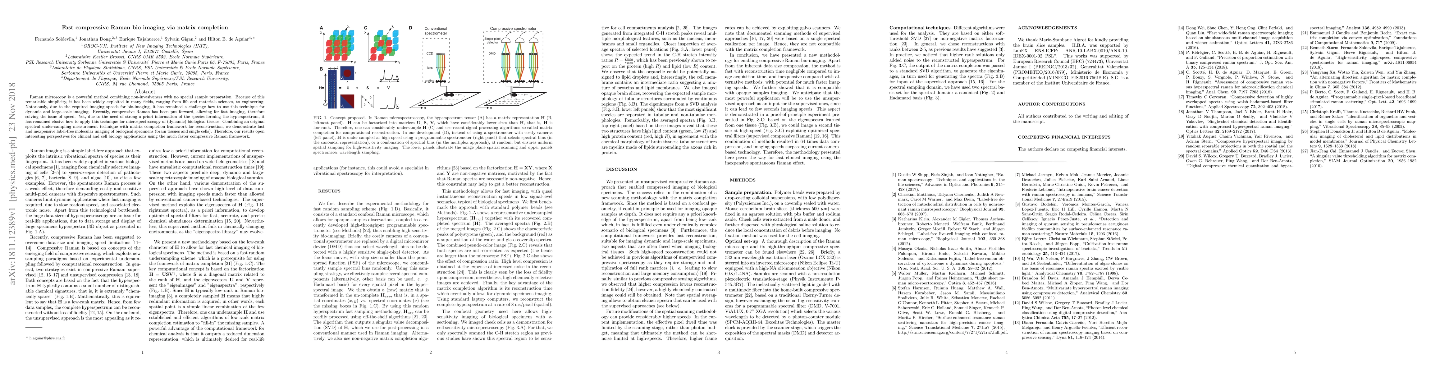
Publication
Metrics
AI Quick Summary
This paper presents a method for fast compressive Raman bio-imaging using matrix completion to reconstruct high-speed, label-free molecular images of biological tissues. The approach enables dynamic and large-scale imaging of brain tissues and single cells, offering potential for clinical and cell biology applications.
Paper Preview
Abstract
Raman microscopy is a powerful method combining non-invasiveness with no special sample preparation. Because of this remarkable simplicity, it has been widely exploited in many fields, ranging from life and materials sciences, to engineering. Notoriously, due to the required imaging speeds for bio-imaging, it has remained a challenge how to use this technique for dynamic and large-scale imaging. Recently, compressive Raman has been put forward, allowing for fast imaging, therefore solving the issue of speed. Yet, due to the need of strong a priori information of the species forming the hyperspectrum, it has remained elusive how to apply this technique for microspectroscopy of (dynamic) biological tissues. Combining an original spectral under-sampling measurement technique with matrix completion framework for reconstruction, we demonstrate fast and inexpensive label-free molecular imaging of biological specimens (brain tissues and single cells). Therefore, our results open interesting perspectives for clinical and cell biology applications using the much faster compressive Raman framework.
AI Key Findings
Get AI-generated insights about this paper's methodology, results, significance, and more — seven facets brought into focus.
Impact
Paper Details
PDF Preview
Key Terms
Citation Network
Current paper (gray), citations (green), references (blue)
Display is limited for performance on very large graphs.
Discussion 0