Publication
Metrics
AI Quick Summary
This paper investigates the rapid formation of hemifused states between free-standing lipid bilayers using an optimized microfluidic technique, achieving formation within hundreds of milliseconds via a dewetting process, significantly faster than previously reported methods. The study demonstrates the stability of these states and their response to an electric field, enhancing understanding of cellular membrane interactions.
Paper Preview
Abstract
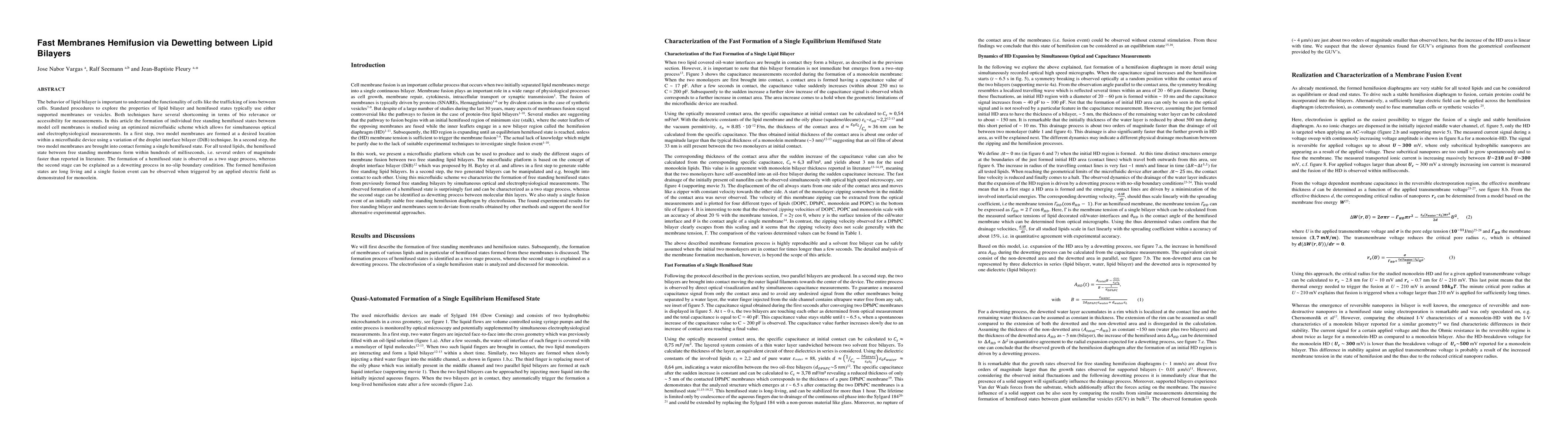
The behavior of lipid bilayer is important to understand the functionality of cells like the trafficking of ions between cells. Standard procedures to explore the properties of lipid bilayer and hemifused states typically use either supported membranes or vesicles. Both techniques have several shortcoming in terms of bio relevance or accessibility for measurements. In this article the formation of individual free standing hemifused states between model cell membranes is studied using an optimized microfluidic scheme which allows for simultaneous optical and electrophysiological measurements. In a first step, two model membranes are formed at a desired location within a microfluidic device using a variation of the droplet interface bilayer (DiB) technique. In a second step, the two model membranes are brought into contact forming a single hemifused state. For all tested lipids, the hemifused state between free standing membranes form within hundreds of milliseconds, i.e. several orders of magnitude faster than reported in literature. The formation of a hemifused state is observed as a two stage process, whereas the second stage can be explained as a dewetting process in no-slip boundary condition. The formed hemifusion states are long living and a single fusion event can be observed when triggered by an applied electric field as demonstrated for monoolein.
AI Key Findings
Get AI-generated insights about this paper's methodology, results, significance, and more — seven facets brought into focus.
Impact
Paper Details
PDF Preview
Key Terms
Citation Network
Current paper (gray), citations (green), references (blue)
Display is limited for performance on very large graphs.
Discussion 0