This paper proposes a fast segmentation method for positron emission tomography (PET) scans using superpixels, principal component analysis, and k-means clustering, achieving an average Dice similarity of 84.2% and significantly reducing computation time by processing fewer superpixels instead of individual pixels.
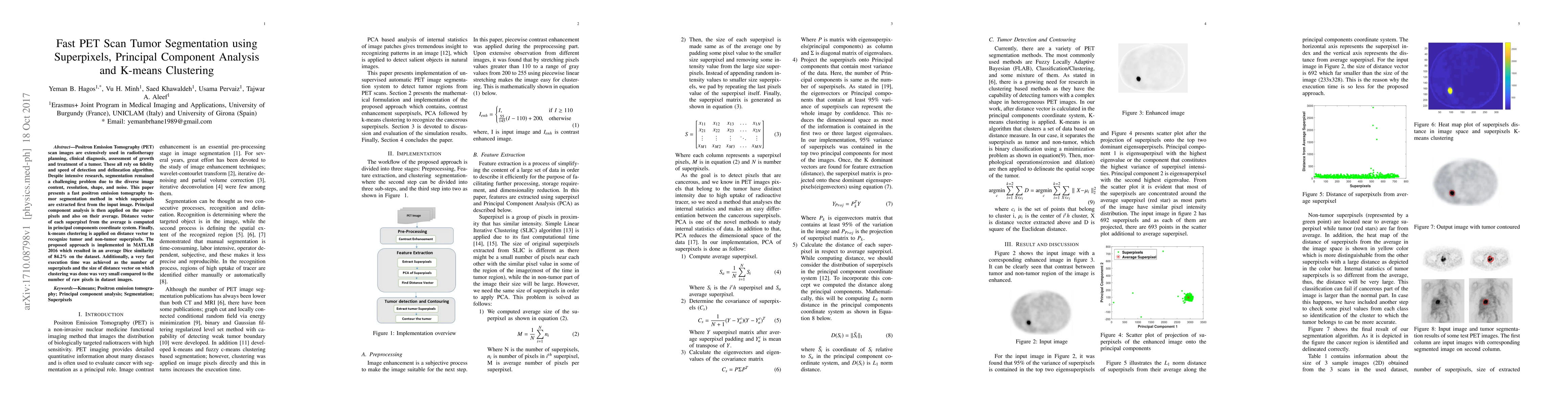
Positron Emission Tomography scan images are extensively used in radiotherapy planning, clinical diagnosis, assessment of growth and treatment of a tumor. These all rely on fidelity and speed of detection and delineation algorithm. Despite intensive research, segmentation remained a challenging problem due to the diverse image content, resolution, shape, and noise. This paper presents a fast positron emission tomography tumor segmentation method in which superpixels are extracted first from the input image. Principal component analysis is then applied on the superpixels and also on their average. Distance vector of each superpixel from the average is computed in principal components coordinate system. Finally, k-means clustering is applied on distance vector to recognize tumor and non-tumor superpixels. The proposed approach is implemented in MATLAB 2016 which resulted in an average Dice similarity of 84.2% on the dataset. Additionally, a very fast execution time was achieved as the number of superpixels and the size of distance vector on which clustering was done was very small compared to the number of raw pixels in dataset images.
Get AI-generated insights about this paper's methodology, results, and significance.
Current paper (gray), citations (green), references (blue)
Title | Authors | Year | Actions |
---|---|---|---|
A New Tool for Extracting Static and Dynamic Parameters from [18F]F-DOPA PET/CT in Pediatric Gliomas
|
Michele Mureddu, T. Funck, G. Morana, Andrea Rossi, A. Ramaglia, C. Milanaccio, A. Verrico, G. Bottoni, Francesco Fiz, A. Piccardo, M. Fato, R. Trò
|
2024
|
View Paper |
Hybrid Clustering-Based Technique to Isolate Tumors in PET/CT Images
|
Enam A. Salman, R. Abdoon, Loay E. George
|
2024
|
View Paper |
Computational Techniques in PET/CT Image Processing for Breast Cancer: A Systematic Mapping Review
|
Karen Carrasco, Lenin Tomalá, Eileen Ramírez Meza, Doris Meza Bolaños, Washington Ramírez Montalvan
|
2024
|
View Paper |
PAPNet: Convolutional network for pancreatic cyst segmentation.
|
Jin Li, Wei Yin, Yuanjun Wang
|
2023
|
View Paper |
Carcinoembryonic Antigens Segmentation and Quantitative Analysis from Fluorescent Images using Principal Component Analysis and Adaptive K-means Clustering
|
M. A. Aslam, Shahzadi Mahnoor, Muhammad Asif Munir, Saman Cheema, Khawaja Humble Hassan, Abdullah Sajid
|
2023
|
View Paper |
Cancerous and Non-Cancerous Brain MRI Classification Method Based on Convolutional Neural Network and Log-Polar Transformation
|
Ferdaus Anam Jibon, Mayeen Uddin Khandaker, M. H. Miraz, Himon Thakur, Fazle Rabby, N. Tamam, A. Sulieman, Y. S. Itas, Hamid Osman
|
2022
|
View Paper |
Artificial intelligence in molecular imaging
|
E. Herskovits
|
2021
|
View Paper |
Pancreatic Tumour Segmentation in Recent Medical Imaging – an Overview
|
A. Sindhu, V. Radha
|
2019
|
View Paper |
Object-Based Convolutional Neural Networks for Cloud and Snow Detection in High-Resolution Multispectral Imagers
|
Lei Wang, Yang Chen, Luliang Tang, Rongshuang Fan, Yunlong Yao
|
2018
|
View Paper |
Multilevel Cloud Detection for High-Resolution Remote Sensing Imagery Using Multiple Convolutional Neural Networks
|
Yang Chen, Rongshuang Fan, M. Bilal, Xiucheng Yang, Jingxue Wang, Wei Li
|
2018
|
View Paper |
Identifying Peat Soil Layers in the South Kalimantan, Indonesia Using K-Means Clustering
|
M. Iryanti, A. Aminudin, E. Agustine, S. Bijaksana, W. Srigutomo, T. Setiawan
|
2018
|
View Paper |
Exploring and comparing data selection methods in the pre-processing step of a deep learning framework for automatic tumor segmentation on PET-CT images
|
E. Korotina
|
View Paper |
Comments (0)