Fast Vascular Ultrasound Imaging with Enhanced Spatial Resolution and Background Rejection
Publication
Metrics
AI Quick Summary
This paper proposes a novel ultrasound imaging technique that applies super-resolution optical fluctuation imaging (SOFI) tools to contrast-enhanced ultrasound (CEUS) plane-wave scans, achieving fast imaging with enhanced spatial resolution and reduced background noise. The method significantly reduces scan duration to less than a second, enabling effective monitoring of vascular flow dynamics and reducing motion artifacts.
Paper Preview
Abstract
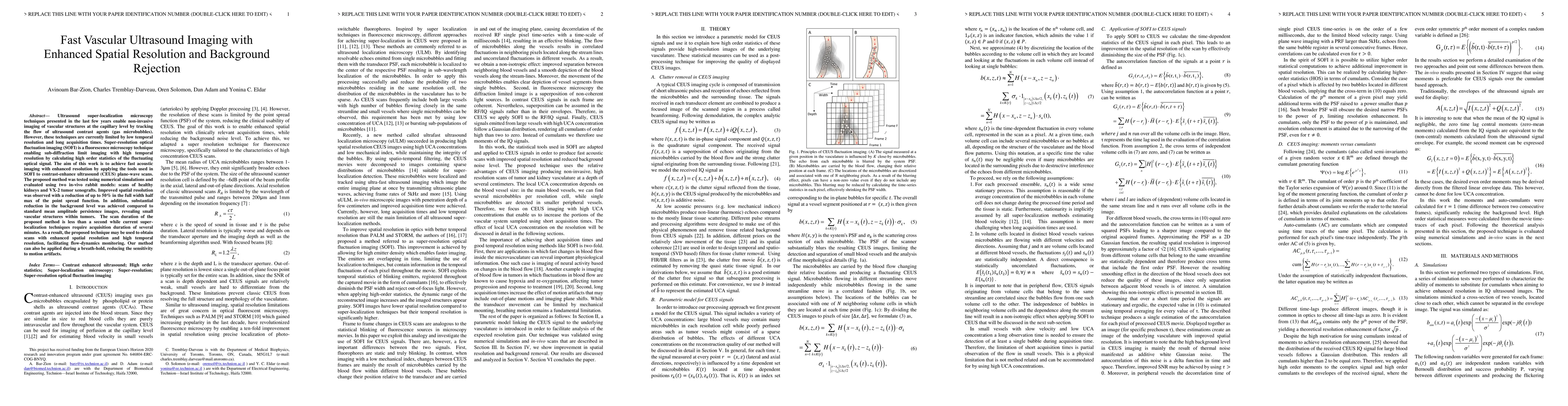
Ultrasound super-localization microscopy techniques presented in the last few years enable non-invasive imaging of vascular structures at the capillary level by tracking the flow of ultrasound contrast agents (gas microbubbles). However, these techniques are currently limited by low temporal resolution and long acquisition times. Super-resolution optical fluctuation imaging (SOFI) is a fluorescence microscopy technique enabling sub-diffraction limit imaging with high temporal resolution by calculating high order statistics of the fluctuating optical signal. The aim of this work is to achieve fast acoustic imaging with enhanced resolution by applying the tools used in SOFI to contrast-enhance ultrasound (CEUS) plane-wave scans. The proposed method was tested using numerical simulations and evaluated using two in-vivo rabbit models: scans of healthy kidneys and VX-2 tumor xenografts. Improved spatial resolution was observed with a reduction of up to 50% in the full width half max of the point spread function. In addition, substantial reduction in the background level was achieved compared to standard mean amplitude persistence images, revealing small vascular structures within tumors. The scan duration of the proposed method is less than a second while current superlocalization techniques require acquisition duration of several minutes. As a result, the proposed technique may be used to obtain scans with enhanced spatial resolution and high temporal resolution, facilitating flow-dynamics monitoring. Our method can also be applied during a breath-hold, reducing the sensitivity to motion artifacts.
AI Key Findings
Get AI-generated insights about this paper's methodology, results, significance, and more — seven facets brought into focus.
Impact
Paper Details
PDF Preview
Key Terms
Citation Network
Current paper (gray), citations (green), references (blue)
Display is limited for performance on very large graphs.
Discussion 0