Publication
Metrics
AI Quick Summary
This study investigates the potential of using muscle fiber orientations from diffusion tensor MRI (DT-MRI) to define the clinical target volume (CTV) boundary for soft-tissue sarcoma treatment. The research utilized MRI data from healthy volunteers to model the CTV boundary, showing promising results in consistency and potential for improving treatment outcomes and reducing amputation rates in sarcoma patients.
Paper Preview
Abstract
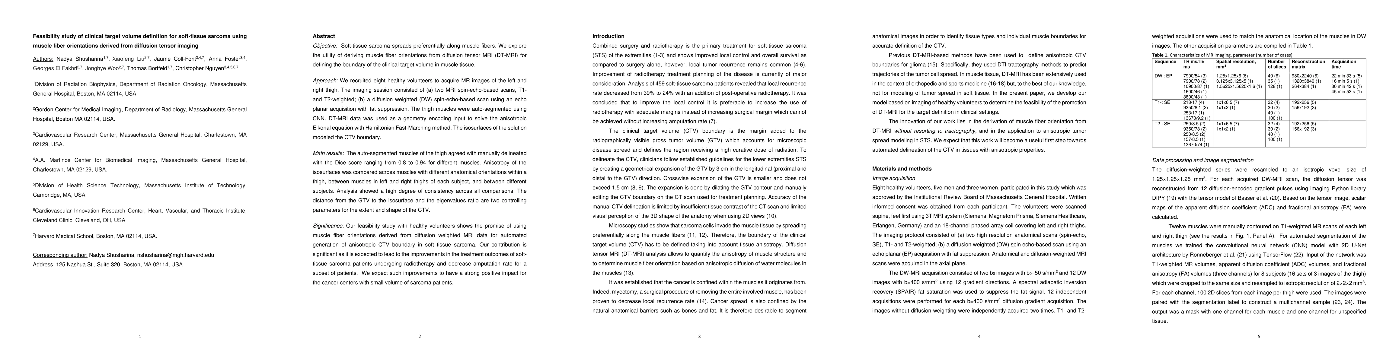
Objective: Soft-tissue sarcoma spreads preferentially along muscle fibers. We explore the utility of deriving muscle fiber orientations from diffusion tensor MRI (DT-MRI) for defining the boundary of the clinical target volume in muscle tissue. Approach: We recruited eight healthy volunteers to acquire MR images of the left and right thigh. The imaging session consisted of (a) two MRI spin-echo-based scans, T1- and T2-weighted; (b) a diffusion weighted (DW) spin-echo-based scan using an echo planar acquisition with fat suppression. The thigh muscles were auto-segmented using CNN. DT-MRI data was used as a geometry encoding input to solve the anisotropic Eikonal equation with Hamiltonian Fast-Marching method. The isosurfaces of the solution modeled the CTV boundary. Main results: The auto-segmented muscles of the thigh agreed with manually delineated with the Dice score ranging from 0.8 to 0.94 for different muscles. Anisotropy of the isosurfaces was compared across muscles with different anatomical orientations within a thigh, between muscles in left and right thighs of each subject, and between different subjects. Analysis showed a high degree of consistency across all comparisons. The distance from the GTV to the isosurface and the eigenvalues ratio are two controlling parameters for the extent and shape of the CTV. Significance: Our feasibility study with healthy volunteers shows the promise of using muscle fiber orientations derived from diffusion weighted MRI data for automated generation of anisotropic CTV boundary in soft tissue sarcoma. Our contribution is significant as it is expected to lead to the improvements in the treatment outcomes of soft-tissue sarcoma patients undergoing radiotherapy and decrease amputation rate for a subset of patients. We expect such improvements to have a strong positive impact for the cancer centers with small volume of sarcoma patients.
AI Key Findings
Get AI-generated insights about this paper's methodology, results, significance, and more — seven facets brought into focus.
Impact
Paper Details
Authors
PDF Preview
Key Terms
Citation Network
Current paper (gray), citations (green), references (blue)
Display is limited for performance on very large graphs.
Discussion 0