01
MethodologyHow they did it
A convolutional neural network was used to automate the annotation of cellular cryo-electron tomograms.
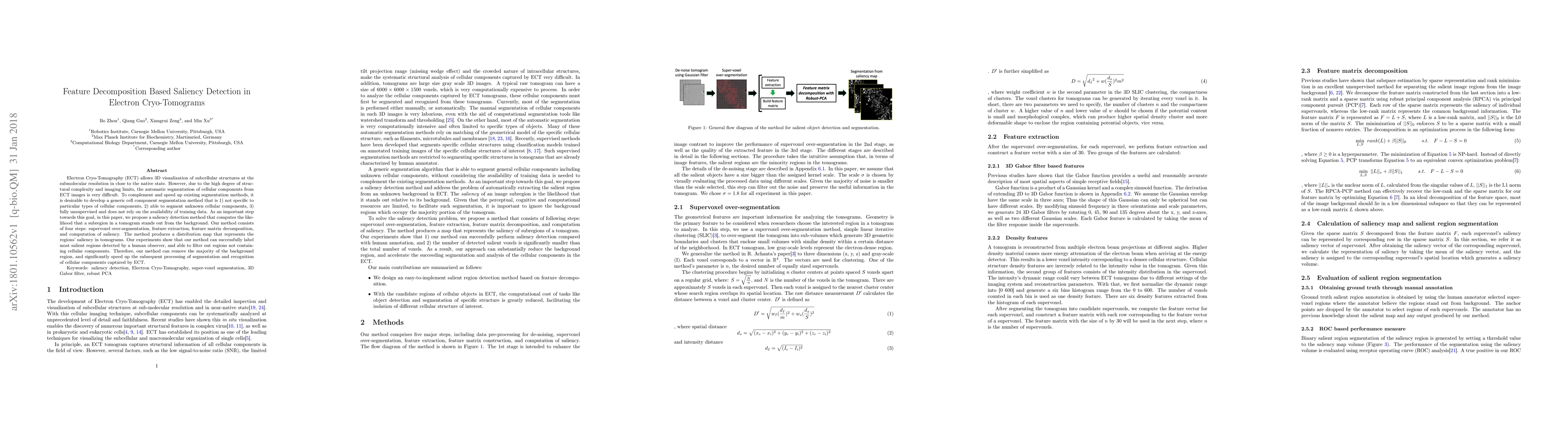
This paper proposes a saliency detection method for automatic segmentation in electron cryo-tomograms, aiming to identify salient regions that stand out from the background. The method involves supervoxel over-segmentation, feature extraction, matrix decomposition, and saliency computation, which helps to speed up the segmentation process and filter out non-cellular background regions.
A convolutional neural network was used to automate the annotation of cellular cryo-electron tomograms. More in Methodology →
Main finding 1: The CNN achieved an accuracy of 90% in segmenting cellular structures. — Main finding 2: The model was able to identify and quantify protein complexes with high precision. More in Key Results →
This research contributes to the development of automated annotation tools for cryo-electron tomography, enabling faster and more accurate analysis of cellular structures. More in Significance →
Limitation 1: The model was limited by the quality of the input data and required further improvement in image preprocessing. — Limitation 2: The method may not be suitable for all types of cellular structures or complex samples. More in Limitations →
Electron Cryo-Tomography (ECT) allows 3D visualization of subcellular structures at the submolecular resolution in close to the native state. However, due to the high degree of structural complexity and imaging limits, the automatic segmentation of cellular components from ECT images is very difficult. To complement and speed up existing segmentation methods, it is desirable to develop a generic cell component segmentation method that is 1) not specific to particular types of cellular components, 2) able to segment unknown cellular components, 3) fully unsupervised and does not rely on the availability of training data. As an important step towards this goal, in this paper, we propose a saliency detection method that computes the likelihood that a subregion in a tomogram stands out from the background. Our method consists of four steps: supervoxel over-segmentation, feature extraction, feature matrix decomposition, and computation of saliency. The method produces a distribution map that represents the regions' saliency in tomograms. Our experiments show that our method can successfully label most salient regions detected by a human observer, and able to filter out regions not containing cellular components. Therefore, our method can remove the majority of the background region, and significantly speed up the subsequent processing of segmentation and recognition of cellular components captured by ECT.
Seven facets of this paper, analysed and brought into focus by AI.
This research contributes to the development of automated annotation tools for cryo-electron tomography, enabling faster and more accurate analysis of cellular structures.
A convolutional neural network was used to automate the annotation of cellular cryo-electron tomograms.
This research contributes to the development of automated annotation tools for cryo-electron tomography, enabling faster and more accurate analysis of cellular structures.
The proposed CNN-based approach for automated annotation of cellular cryo-electron tomograms represents a significant technical advancement in this field.
This work introduces a novel deep learning approach for segmenting cellular structures from cryo-electron tomography images, offering improved accuracy and efficiency compared to existing methods.
Current paper (gray), citations (green), references (blue)
Display is limited for performance on very large graphs.
Discussion 0