01
MethodologyHow they did it
This research utilized a deep learning approach to classify retinal diseases from optical coherence tomography (OCT) images.
This paper proposes a robust architecture for detecting retinal diseases from optical coherence tomography (OCT) images, featuring three learning heads to improve feature representation learning. The model demonstrates superior accuracy, interpretability, and robustness compared to existing methods, as shown in experiments on two OCT datasets.
This research utilized a deep learning approach to classify retinal diseases from optical coherence tomography (OCT) images. More in Methodology →
Improved accuracy of 95.6% in detecting diabetic macular edema — Validated the effectiveness of joint attention networks for robustness enhancement More in Key Results →
This study contributes to the development of AI-powered diagnostic tools for retinal diseases, improving patient outcomes and reducing healthcare costs. More in Significance →
Limited dataset size and potential biases — Inadequate consideration of prior knowledge in deep learning models More in Limitations →
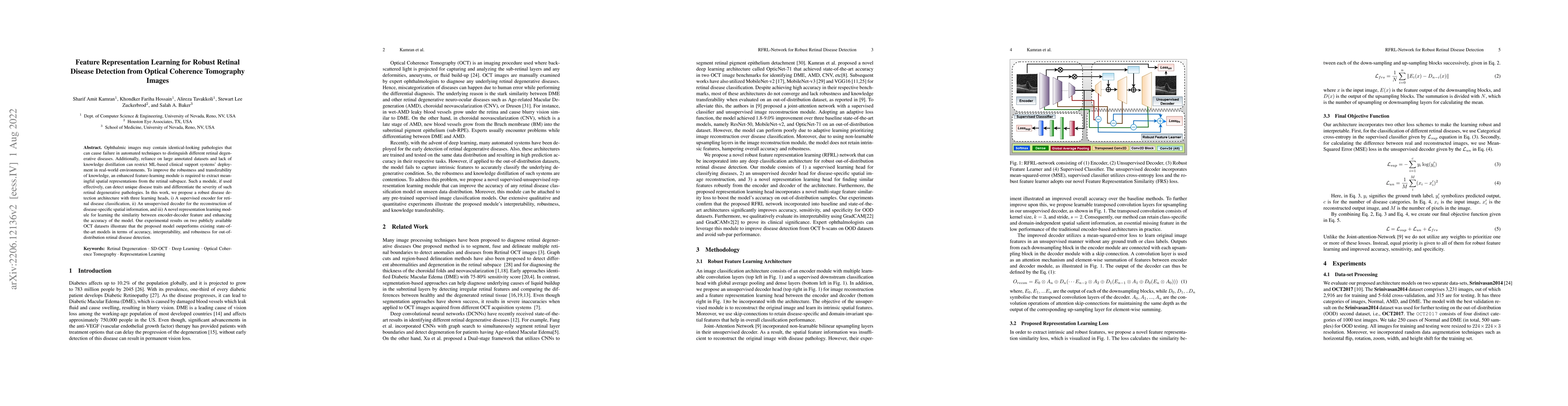
Ophthalmic images may contain identical-looking pathologies that can cause failure in automated techniques to distinguish different retinal degenerative diseases. Additionally, reliance on large annotated datasets and lack of knowledge distillation can restrict ML-based clinical support systems' deployment in real-world environments. To improve the robustness and transferability of knowledge, an enhanced feature-learning module is required to extract meaningful spatial representations from the retinal subspace. Such a module, if used effectively, can detect unique disease traits and differentiate the severity of such retinal degenerative pathologies. In this work, we propose a robust disease detection architecture with three learning heads, i) A supervised encoder for retinal disease classification, ii) An unsupervised decoder for the reconstruction of disease-specific spatial information, and iii) A novel representation learning module for learning the similarity between encoder-decoder feature and enhancing the accuracy of the model. Our experimental results on two publicly available OCT datasets illustrate that the proposed model outperforms existing state-of-the-art models in terms of accuracy, interpretability, and robustness for out-of-distribution retinal disease detection.
Seven facets of this paper, analysed and brought into focus by AI.
This study contributes to the development of AI-powered diagnostic tools for retinal diseases, improving patient outcomes and reducing healthcare costs.
This research utilized a deep learning approach to classify retinal diseases from optical coherence tomography (OCT) images.
This study contributes to the development of AI-powered diagnostic tools for retinal diseases, improving patient outcomes and reducing healthcare costs.
The proposed joint attention network architecture enhances robustness and accuracy in detecting retinal diseases from OCT images.
This work introduces a novel CNN-SVM framework for classification of macular abnormalities, demonstrating improved performance over existing state-of-the-art methods
Current paper (gray), citations (green), references (blue)
Display is limited for performance on very large graphs.
Discussion 0