Publication
Metrics
AI Quick Summary
This paper demonstrates femtosecond mega-electron-volt electron microdiffraction achieving 5 μm beam size and 100 fs temporal resolution, enabling visualization of ultrafast structural changes in materials. High-quality diffraction from paraffin and phonon softening in Bi showcases the technique's potential for advancing material and biological sciences.
Paper Preview
Abstract
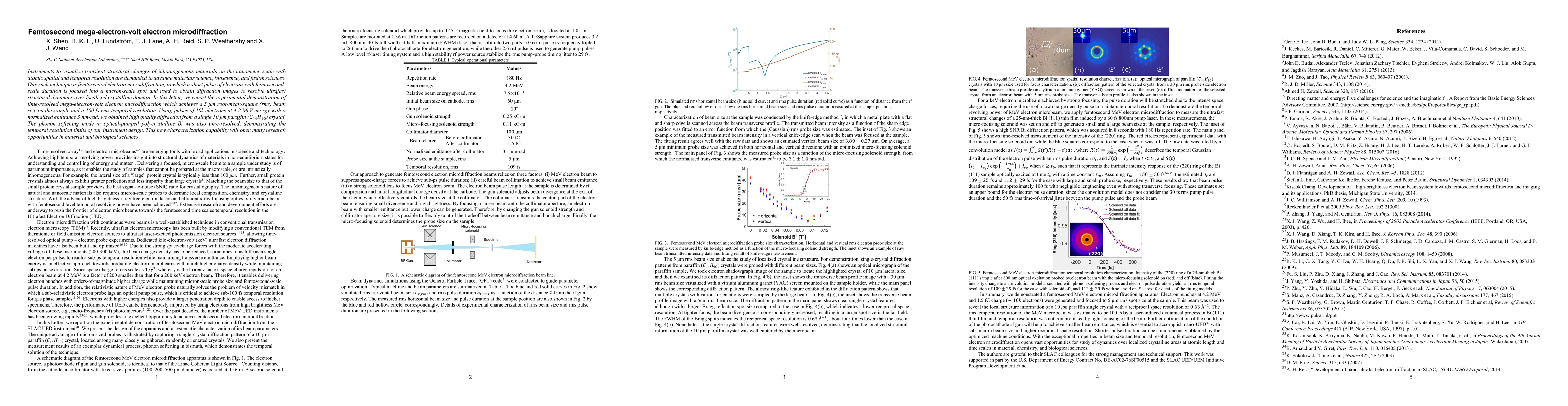
Instruments to visualize transient structural changes of inhomogeneous materials on the nanometer scale with atomic spatial and temporal resolution are demanded to advance materials science, bioscience, and fusion sciences. One such technique is femtosecond electron microdiffraction, in which a short pulse of electrons with femtosecond-scale duration is focused into a micron-scale spot and used to obtain diffraction images to resolve ultrafast structural dynamics over localized crystalline domain. In this letter, we report the experimental demonstration of time-resolved mega-electron-volt electron microdiffraction which achieves a 5 {\mu}m root-mean-square (rms) beam size on the sample and a 100 fs rms temporal resolution. Using pulses of 10k electrons at 4.2 MeV energy with a normalized emittance 3 nm-rad, we obtained high quality diffraction from a single 10 {\mu}m paraffin (C_44 H_90) crystal. The phonon softening mode in optical-pumped polycrystalline Bi was also time-resolved, demonstrating the temporal resolution limits of our instrument design. This new characterization capability will open many research opportunities in material and biological sciences.
AI Key Findings
Get AI-generated insights about this paper's methodology, results, significance, and more — seven facets brought into focus.
Impact
Paper Details
PDF Preview
Key Terms
Citation Network
Current paper (gray), citations (green), references (blue)
Display is limited for performance on very large graphs.
Discussion 0