Publication
Metrics
AI Quick Summary
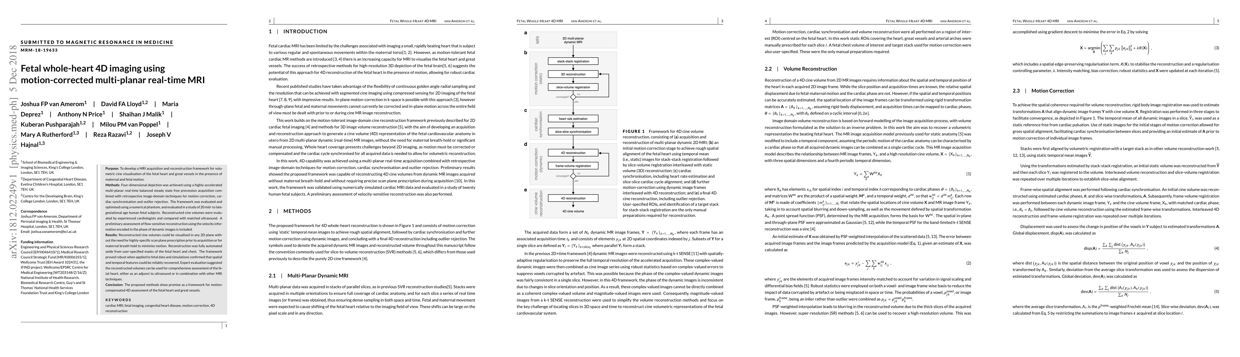
This paper presents a novel MRI framework for 4D volumetric cine imaging of the fetal heart, using accelerated multi-planar real-time acquisition and retrospective motion correction. The method enables comprehensive fetal heart assessment without requiring specific scan planes or maternal breath holds, showing promise as an adjunct to ultrasound or combined with other MRI techniques.
Paper Preview
Abstract
Purpose: To develop a MRI acquisition and reconstruction framework for volumetric cine visualisation of the fetal heart and great vessels in the presence of maternal and fetal motion. Methods: Four-dimensional depiction was achieved using a highly-accelerated multi-planar real-time balanced steady state free precession acquisition combined with retrospective image-domain techniques for motion correction, cardiac synchronisation and outlier rejection. The framework was evaluated and optimised using a numerical phantom, and evaluated in a study of 20 mid- to late-gestational age human fetal subjects. Reconstructed cine volumes were evaluated by experienced cardiologists and compared with matched ultrasound. A preliminary assessment of flow-sensitive reconstruction using the velocity information encoded in the phase of dynamic images is included. Results: Reconstructed cine volumes could be visualised in any 2D plane without the need for highly-specific scan plane prescription prior to acquisition or for maternal breath hold to minimise motion. Reconstruction was fully automated aside from user-specified masks of the fetal heart and chest. The framework proved robust when applied to fetal data and simulations confirmed that spatial and temporal features could be reliably recovered. Expert evaluation suggested the reconstructed volumes can be used for comprehensive assessment of the fetal heart, either as an adjunct to ultrasound or in combination with other MRI techniques. Conclusion: The proposed methods show promise as a framework for motion-compensated 4D assessment of the fetal heart and great vessels.
AI Key Findings
Get AI-generated insights about this paper's methodology, results, significance, and more — seven facets brought into focus.
Impact
Paper Details
PDF Preview
Key Terms
Citation Network
Current paper (gray), citations (green), references (blue)
Display is limited for performance on very large graphs.
Discussion 0