Publication
Metrics
Quick Actions
AI Quick Summary
FGR-Net is a novel framework that combines an autoencoder with a classifier to automatically assess the quality of retinal fundus images, distinguishing between gradable and non-gradable images. The autoencoder reconstructs the input image to extract visual features, which are then classified, while interpretability methods provide visual feedback for clinicians. Experimental results show FGR-Net's superior performance with 89% accuracy and 87% F1-score.
Quick Answers
What is "FGR-Net:Interpretable fundus imagegradeability classification based on deepreconstruction learning" about?
FGR-Net is a novel framework that combines an autoencoder with a classifier to automatically assess the quality of retinal fundus images, distinguishing between gradable and non-gradable images. The autoencoder reconstructs the input image to extract visual features, which are then classified, while interpretability methods provide visual feedback for clinicians. Experimental results show FGR-Net's superior performance with 89% accuracy and 87% F1-score.
What methodology did the authors use?
The research introduces FGR-Net, a novel framework that combines an autoencoder network with a classifier network to automatically assess and interpret underlying fundus image quality. FGR-Net uses a deep autoencoder for self-supervised learning to extract visual characteristics from input fundus images, which are then fed into a deep classifier network to distinguish between gradable and ungradable images. More in Methodology →
What are the key results?
FGR-Net achieved an accuracy of 89% and an F1-score of 87% in classifying fundus image gradeability. — The autoencoder in FGR-Net was found to be a key factor in forcing the classifier to focus on relevant structures like the fovea, optic disk, and prominent blood vessels. More in Key Results →
Why is this work significant?
This research is significant as it presents a novel framework (FGR-Net) that not only classifies fundus image gradeability but also provides interpretable quality assessment, which is crucial for the performance of diagnostic CAD systems for retinal diseases. More in Significance →
What are the main limitations?
The study did not investigate the relationship between model accuracy and the visualization quality of interpretability methods for distinguishing gradable and non-gradable retinal images. — The performance evaluation was conducted on specific datasets (EyePACS, Eye-Quality, and an in-house dataset from HUSJR, Spain) and may not generalize to all types of fundus images. More in Limitations →
Abstract
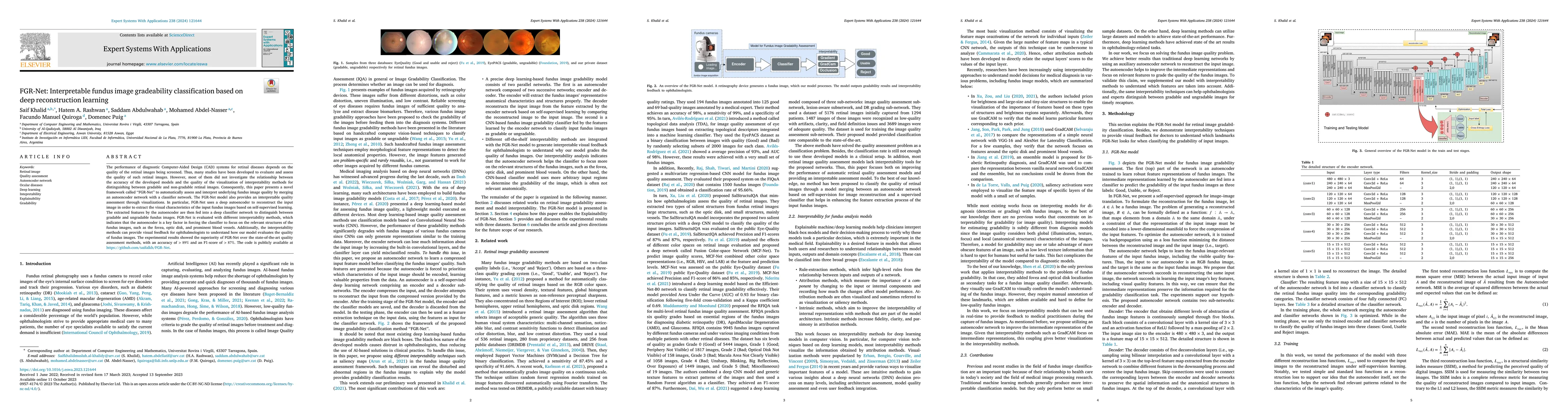
The performance of diagnostic Computer-Aided Design (CAD) systems for retinal diseases depends on the quality of the retinal images being screened. Thus, many studies have been developed to evaluate and assess the quality of such retinal images. However, most of them did not investigate the relationship between the accuracy of the developed models and the quality of the visualization of interpretability methods for distinguishing between gradable and non-gradable retinal images. Consequently, this paper presents a novel framework called FGR-Net to automatically assess and interpret underlying fundus image quality by merging an autoencoder network with a classifier network. The FGR-Net model also provides an interpretable quality assessment through visualizations. In particular, FGR-Net uses a deep autoencoder to reconstruct the input image in order to extract the visual characteristics of the input fundus images based on self-supervised learning. The extracted features by the autoencoder are then fed into a deep classifier network to distinguish between gradable and ungradable fundus images. FGR-Net is evaluated with different interpretability methods, which indicates that the autoencoder is a key factor in forcing the classifier to focus on the relevant structures of the fundus images, such as the fovea, optic disk, and prominent blood vessels. Additionally, the interpretability methods can provide visual feedback for ophthalmologists to understand how our model evaluates the quality of fundus images. The experimental results showed the superiority of FGR-Net over the state-of-the-art quality assessment methods, with an accuracy of 89% and an F1-score of 87%.
AI Key Findings
Generated Sep 01, 2025
Methodology — What approach did the authors take?
The research introduces FGR-Net, a novel framework that combines an autoencoder network with a classifier network to automatically assess and interpret underlying fundus image quality. FGR-Net uses a deep autoencoder for self-supervised learning to extract visual characteristics from input fundus images, which are then fed into a deep classifier network to distinguish between gradable and ungradable images.
Key Results — What are the main findings?
- FGR-Net achieved an accuracy of 89% and an F1-score of 87% in classifying fundus image gradeability.
- The autoencoder in FGR-Net was found to be a key factor in forcing the classifier to focus on relevant structures like the fovea, optic disk, and prominent blood vessels.
- Experimental results demonstrated the superiority of FGR-Net over state-of-the-art quality assessment methods.
- Interpretability methods provided visual feedback for ophthalmologists to understand how FGR-Net evaluates fundus image quality.
Significance — Why does this research matter?
This research is significant as it presents a novel framework (FGR-Net) that not only classifies fundus image gradeability but also provides interpretable quality assessment, which is crucial for the performance of diagnostic CAD systems for retinal diseases.
Technical Contribution — What is the technical contribution?
The technical contribution of this work is the development of FGR-Net, a framework that merges deep autoencoder reconstruction learning with a classifier network for interpretable fundus image gradeability classification.
Novelty — What is new about this work?
FGR-Net stands out by providing an interpretable quality assessment for fundus images, focusing on relevant structures through the autoencoder, and offering visual feedback for ophthalmologists, which distinguishes it from existing quality assessment methods.
Limitations — What are the limitations of this study?
- The study did not investigate the relationship between model accuracy and the visualization quality of interpretability methods for distinguishing gradable and non-gradable retinal images.
- The performance evaluation was conducted on specific datasets (EyePACS, Eye-Quality, and an in-house dataset from HUSJR, Spain) and may not generalize to all types of fundus images.
Future Work — What did the authors propose for future work?
- Further research could explore the application of FGR-Net on larger and more diverse datasets to validate its generalizability.
- Investigating the integration of FGR-Net with existing CAD systems for retinal diseases to assess its impact on diagnostic accuracy.
How to Cite This Paper
@article{puig2024fgr,
title = {FGR-Net:Interpretable fundus imagegradeability classification based on
deepreconstruction learning},
author = {Puig, Domenec and Quiroga, Facundo Manuel and Rashwan, Hatem A. and others},
year = {2024},
eprint = {2409.10246},
archivePrefix = {arXiv},
primaryClass = {eess.IV},
}
Puig, D., Quiroga, F., Rashwan, H., Abdel-Nasser, M., Abdulwahab, S., & Khalid, S. (2024). FGR-Net:Interpretable fundus imagegradeability classification based on
deepreconstruction learning. arXiv. https://arxiv.org/abs/2409.10246
Puig, Domenec, et al. "FGR-Net:Interpretable fundus imagegradeability classification based on
deepreconstruction learning." arXiv, 2024, arxiv.org/abs/2409.10246.
PDF Preview
Citation Network
Current paper (gray), citations (green), references (blue)
Display is limited for performance on very large graphs.
Similar Papers
Found 4 papersDeep learning-based classification of multiple fundus diseases using ultra-widefield images.
Tu, Xiang, Duan, Ming-Ming
Correction: Resilient back-propagation machine learning-based classification on fundus images for retinal microaneurysm detection.
Steffi, S
A deep learning model for classification of diabetic retinopathy in eye fundus images based on retinal lesion detection
Oscar J. Perdomo, Melissa delaPava, Hernán Ríos et al.
| Title | Authors | Year | Actions |
|---|
Comments (0)