Quick Answers

What methodology did the authors use?

The study employed a two-step transfer learning approach combining patch-level and slide-level classifiers. Pixel-wise annotations were created by expert pathologists, and a phase correlation-based image registration algorithm was used to align annotations across different scanner versions of the same slide. More in Methodology →

What are the key results?

The model achieved a mean pairwise Cohen's kappa of 0.66 with pathologists on the STHLM3 cohort — Cross-scanner reproducibility showed high agreement with Cohen's kappa values ranging from 0.90 to 0.97 More in Key Results →

Why is this work significant?

This research provides a robust AI solution for cribriform morphology detection in prostate cancer histopathology, improving diagnostic consistency and enabling cross-scanner compatibility More in Significance →

What are the main limitations?

The model's performance is dependent on high-quality pixel-wise annotations — The study primarily used Swedish cohorts which may limit generalizability More in Limitations →

Paper Preview

Abstract

Background: Cribriform morphology in prostate cancer is a histological feature that indicates poor prognosis and contraindicates active surveillance. However, it remains underreported and subject to significant interobserver variability amongst pathologists. We aimed to develop and validate an AI-based system to improve cribriform pattern detection. 
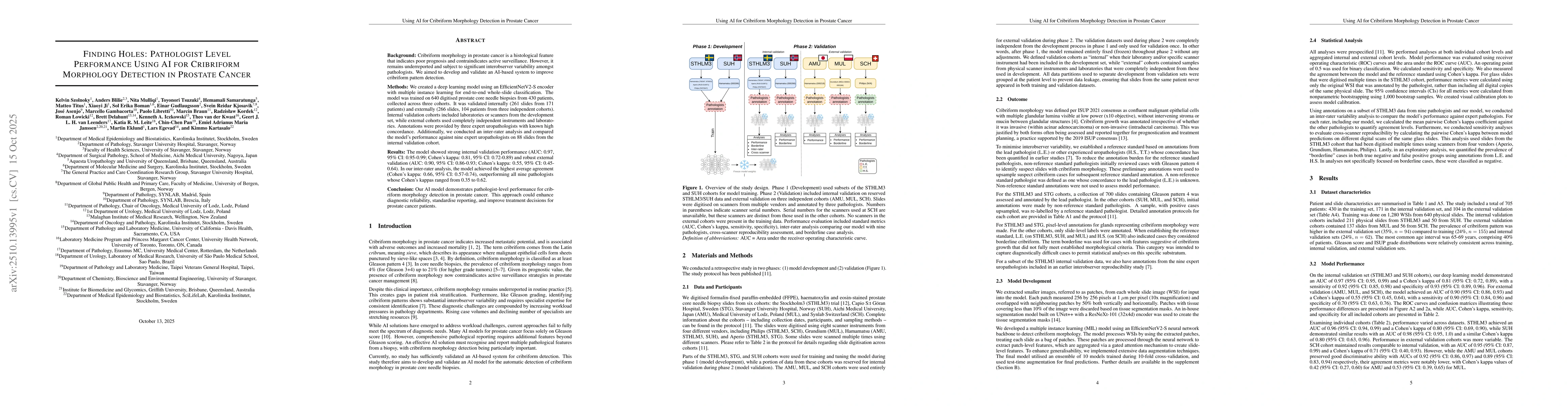
Methods: We created a deep learning model using an EfficientNetV2-S encoder with multiple instance learning for end-to-end whole-slide classification. The model was trained on 640 digitised prostate core needle biopsies from 430 patients, collected across three cohorts. It was validated internally (261 slides from 171 patients) and externally (266 slides, 104 patients from three independent cohorts). Internal validation cohorts included laboratories or scanners from the development set, while external cohorts used completely independent instruments and laboratories. Annotations were provided by three expert uropathologists with known high concordance. Additionally, we conducted an inter-rater analysis and compared the model's performance against nine expert uropathologists on 88 slides from the internal validation cohort. Results: The model showed strong internal validation performance (AUC: 0.97, 95% CI: 0.95-0.99; Cohen's kappa: 0.81, 95% CI: 0.72-0.89) and robust external validation (AUC: 0.90, 95% CI: 0.86-0.93; Cohen's kappa: 0.55, 95% CI: 0.45-0.64). In our inter-rater analysis, the model achieved the highest average agreement (Cohen's kappa: 0.66, 95% CI: 0.57-0.74), outperforming all nine pathologists whose Cohen's kappas ranged from 0.35 to 0.62. Conclusion: Our AI model demonstrates pathologist-level performance for cribriform morphology detection in prostate cancer. This approach could enhance diagnostic reliability, standardise reporting, and improve treatment decisions for prostate cancer patients.

AI Key Findings

Generated Oct 30, 2025

Methodology — What approach did the authors take?

The study employed a two-step transfer learning approach combining patch-level and slide-level classifiers. Pixel-wise annotations were created by expert pathologists, and a phase correlation-based image registration algorithm was used to align annotations across different scanner versions of the same slide.

Key Results — What are the main findings?

  • The model achieved a mean pairwise Cohen's kappa of 0.66 with pathologists on the STHLM3 cohort
  • Cross-scanner reproducibility showed high agreement with Cohen's kappa values ranging from 0.90 to 0.97
  • The model demonstrated good calibration across different cohorts

Significance — Why does this research matter?

This research provides a robust AI solution for cribriform morphology detection in prostate cancer histopathology, improving diagnostic consistency and enabling cross-scanner compatibility

Technical Contribution — What is the technical contribution?

Implementation of a two-step transfer learning framework with gated attention mechanism for slide-level feature aggregation

Novelty — What is new about this work?

First application of phase correlation-based image registration for aligning annotations across different scanner versions of the same histopathological slide

Limitations — What are the limitations of this study?

  • The model's performance is dependent on high-quality pixel-wise annotations
  • The study primarily used Swedish cohorts which may limit generalizability

Future Work — What did the authors propose for future work?

  • Developing multi-center validation studies with diverse cohorts
  • Improving model performance on borderline cases

Paper Details

Paper ID: 2510.13995
Categories:
cs.CV cs.AI

How to Cite This Paper

BibTeX
@article{blilie2025finding,
  title         = {Finding Holes: Pathologist Level Performance Using AI for Cribriform
  Morphology Detection in Prostate Cancer},
  author        = {Blilie, Anders and Delahunt, Brett and Pan, Chin-Chen and others},
  year          = {2025},
  eprint        = {2510.13995},
  archivePrefix = {arXiv},
  primaryClass  = {cs.CV},
}
APA 7
Blilie, A., Delahunt, B., Pan, C., Gudlaugsson, E., Janssen, E., Leenders, G., Samaratunga, H., Asenjo, J., Leite, K., Szolnoky, K., Iczkowski, K., Kartasalo, K., Egevad, L., Gambacorta, M., Braun, M., Eklund, M., Titus, M., Mulliqi, N., Libretti, P., Kordek, R., Łowicki, R., Boman, S., Kjosavik, S., Kwast, T., Tsuzuki, T., & Ji, X. (2025). Finding Holes: Pathologist Level Performance Using AI for Cribriform
  Morphology Detection in Prostate Cancer. arXiv. https://arxiv.org/abs/2510.13995
MLA 9
Blilie, Anders, et al. "Finding Holes: Pathologist Level Performance Using AI for Cribriform
  Morphology Detection in Prostate Cancer." arXiv, 2025, arxiv.org/abs/2510.13995.

Similar Papers

Found 4 papers

Pathologist-like explainable AI for interpretable Gleason grading in prostate cancer

Gesa Mittmann, Thomas Kalinski, Christian Hörner et al.

2024-10-19

Cribriform-Positive and -Negative Prostate Cancer.

D'Amico, Anthony V, Minhas, Paras S

2026-02-26

Cribriform-Positive and -Negative Prostate Cancer-Reply.

Sushentsev, Nikita, Warren, Anne Y, Hamdy, Freddie C

2026-02-26

Cribriform pattern detection in prostate histopathological images using deep learning models

Chin Fong Wong, Danilo Medina Giron, Emarene Mationg Kalaw et al.

2019-10-10

Comments (0)

This website uses cookies to ensure you get the best experience. Learn more