Finding Nano-\"Otzi: Semi-Supervised Volume Visualization for Cryo-Electron Tomography
Publication
Metrics
AI Quick Summary
This paper proposes a semi-supervised visualization method for cryo-electron tomography data, leveraging soft segmentation and combining weak and deep-learning based segmentation algorithms to enhance volume visualization in the presence of low signal-to-noise ratios. The technique uses gradient-free ambient occlusion shading to highlight structural details while suppressing noise, demonstrated effectively on SARS-CoV-2 virions.
Paper Preview
Abstract
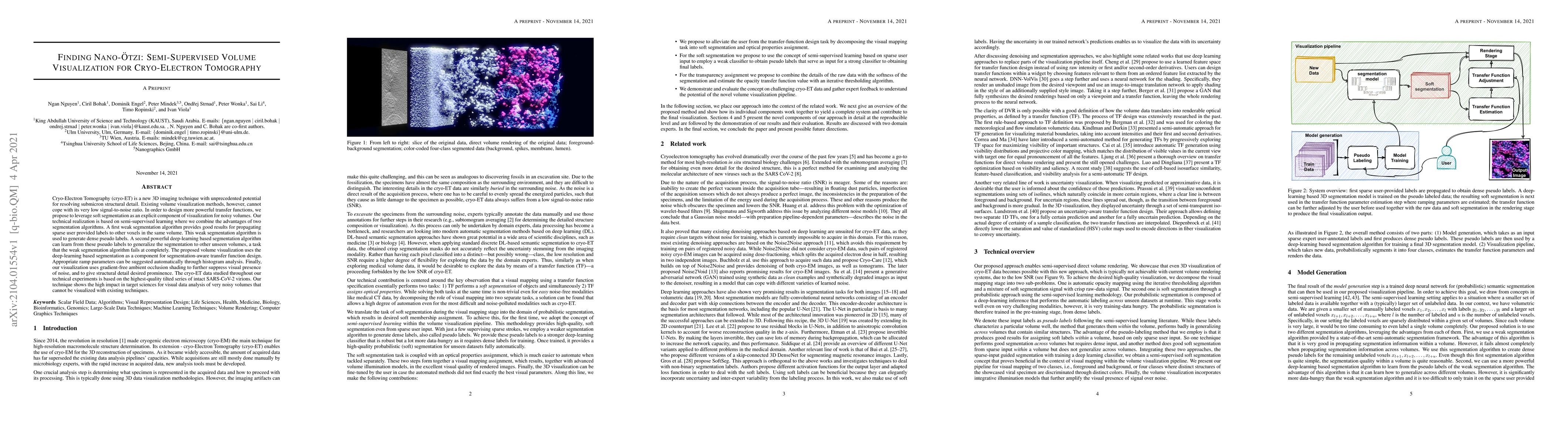
Cryo-Electron Tomography (cryo-ET) is a new 3D imaging technique with unprecedented potential for resolving submicron structural detail. Existing volume visualization methods, however, cannot cope with its very low signal-to-noise ratio. In order to design more powerful transfer functions, we propose to leverage soft segmentation as an explicit component of visualization for noisy volumes. Our technical realization is based on semi-supervised learning where we combine the advantages of two segmentation algorithms. A first weak segmentation algorithm provides good results for propagating sparse user provided labels to other voxels in the same volume. This weak segmentation algorithm is used to generate dense pseudo labels. A second powerful deep-learning based segmentation algorithm can learn from these pseudo labels to generalize the segmentation to other unseen volumes, a task that the weak segmentation algorithm fails at completely. The proposed volume visualization uses the deep-learning based segmentation as a component for segmentation-aware transfer function design. Appropriate ramp parameters can be suggested automatically through histogram analysis. Finally, our visualization uses gradient-free ambient occlusion shading to further suppress visual presence of noise, and to give structural detail desired prominence. The cryo-ET data studied throughout our technical experiments is based on the highest-quality tilted series of intact SARS-CoV-2 virions. Our technique shows the high impact in target sciences for visual data analysis of very noisy volumes that cannot be visualized with existing techniques.
AI Key Findings
Get AI-generated insights about this paper's methodology, results, significance, and more — seven facets brought into focus.
Impact
Paper Details
Authors
PDF Preview
Key Terms
Citation Network
Current paper (gray), citations (green), references (blue)
Display is limited for performance on very large graphs.
Discussion 0