Quick Summary:

This paper introduces a method to improve the interpretability of Diabetic Retinopathy staging using graph-based knowledge and vision-language models. It constructs biologically informed graphs from OCTA images to encode retinal features, employs a graph neural network for staging, and uses integrated gradients for explainability. The approach enhances both classification accuracy and provides clinically interpretable diagnostic explanations.

Summary

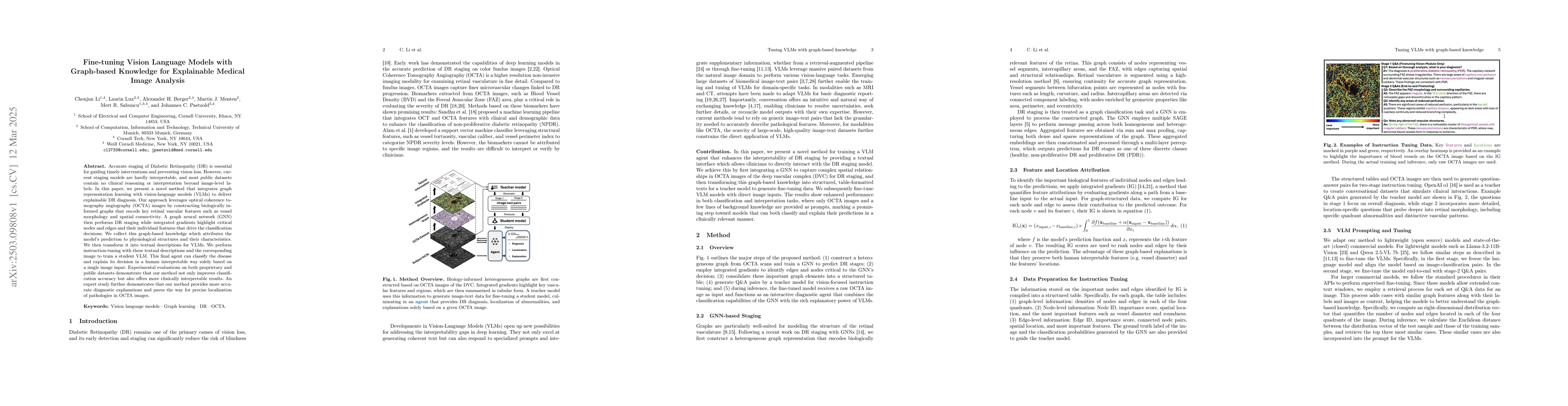
Accurate staging of Diabetic Retinopathy (DR) is essential for guiding timely interventions and preventing vision loss. However, current staging models are hardly interpretable, and most public datasets contain no clinical reasoning or interpretation beyond image-level labels. In this paper, we present a novel method that integrates graph representation learning with vision-language models (VLMs) to deliver explainable DR diagnosis. Our approach leverages optical coherence tomography angiography (OCTA) images by constructing biologically informed graphs that encode key retinal vascular features such as vessel morphology and spatial connectivity. A graph neural network (GNN) then performs DR staging while integrated gradients highlight critical nodes and edges and their individual features that drive the classification decisions. We collect this graph-based knowledge which attributes the model's prediction to physiological structures and their characteristics. We then transform it into textual descriptions for VLMs. We perform instruction-tuning with these textual descriptions and the corresponding image to train a student VLM. This final agent can classify the disease and explain its decision in a human interpretable way solely based on a single image input. Experimental evaluations on both proprietary and public datasets demonstrate that our method not only improves classification accuracy but also offers more clinically interpretable results. An expert study further demonstrates that our method provides more accurate diagnostic explanations and paves the way for precise localization of pathologies in OCTA images.

AI Key Findings

Generated Jun 10, 2025

Methodology

The research integrates graph representation learning with vision-language models (VLMs) for explainable Diabetic Retinopathy (DR) diagnosis using optical coherence tomography angiography (OCTA) images. Biologically informed graphs encode vascular features, and a graph neural network (GNN) performs DR staging. Integrated gradients highlight critical nodes and edges driving classification decisions, which are then transformed into textual descriptions for VLMs through instruction-tuning.

Key Results

  • The proposed method improves classification accuracy for DR staging.
  • The method offers more clinically interpretable results compared to baseline models.
  • Experimental evaluations on proprietary and public datasets demonstrate the effectiveness of the approach.
  • Expert study confirms that the method provides more accurate diagnostic explanations and aids in precise localization of pathologies in OCTA images.
  • Graph-knowledge-fine-tuned (GFT) models outperform baseline vision-language models and standard fine-tuning approaches on proprietary datasets.

Significance

This research is significant as it addresses the lack of interpretability in current DR staging models, which is crucial for guiding timely interventions and preventing vision loss. The method offers a novel approach to integrate graph-based knowledge with VLMs, enabling explainable medical image analysis.

Technical Contribution

The main technical contribution is the integration of graph-based knowledge with VLMs for explainable medical image analysis, specifically for DR diagnosis using OCTA images.

Novelty

This work is novel by combining graph representation learning with vision-language models for explainable medical image analysis, focusing on DR diagnosis. It introduces a method to translate complex vascular patterns into structured textual descriptions for effective instruction-tuning.

Limitations

  • The paper does not explicitly discuss limitations.
  • Potential limitations could include reliance on high-quality OCTA images and the need for specialized knowledge to interpret graph-based explanations.

Future Work

  • Exploration of the method's potential in clinical settings.
  • Investigation of synthetic image generation to augment training data and enhance model robustness.

Paper Details

PDF Preview

Download PDF

Key Terms

No key terms available for this paper.

Citation Network

Current paper (gray), citations (green), references (blue)

Current Paper
Citations
References
Click to view

No citations found for this paper.

Comments (0)

This website uses cookies to ensure you get the best experience. Learn more