Finite-Difference Time-Domain Simulation for Three-dimensional Polarized Light Imaging
Publication
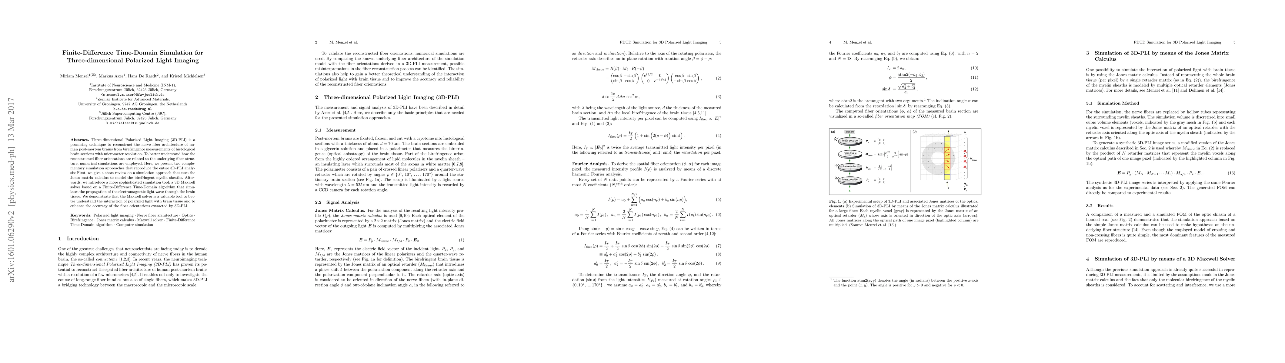
Metrics
AI Quick Summary
This paper presents two simulation methods for 3D Polarized Light Imaging (3D-PLI) to reconstruct nerve fiber architecture in human brains. The first uses Jones matrix calculus for birefringence modeling, while the second employs a sophisticated 3D Maxwell solver based on Finite-Difference Time-Domain to simulate light wave propagation, enhancing the accuracy of fiber orientation extraction.
Paper Preview
Abstract
Three-dimensional Polarized Light Imaging (3D-PLI) is a promising technique to reconstruct the nerve fiber architecture of human post-mortem brains from birefringence measurements of histological brain sections with micrometer resolution. To better understand how the reconstructed fiber orientations are related to the underlying fiber structure, numerical simulations are employed. Here, we present two complementary simulation approaches that reproduce the entire 3D-PLI analysis: First, we give a short review on a simulation approach that uses the Jones matrix calculus to model the birefringent myelin sheaths. Afterwards, we introduce a more sophisticated simulation tool: a 3D Maxwell solver based on a Finite-Difference Time-Domain algorithm that simulates the propagation of the electromagnetic light wave through the brain tissue. We demonstrate that the Maxwell solver is a valuable tool to better understand the interaction of polarized light with brain tissue and to enhance the accuracy of the fiber orientations extracted by 3D-PLI.
AI Key Findings
Get AI-generated insights about this paper's methodology, results, significance, and more — seven facets brought into focus.
Impact
Paper Details
PDF Preview
Key Terms
Citation Network
Current paper (gray), citations (green), references (blue)
Display is limited for performance on very large graphs.
Discussion 0