01
MethodologyHow they did it
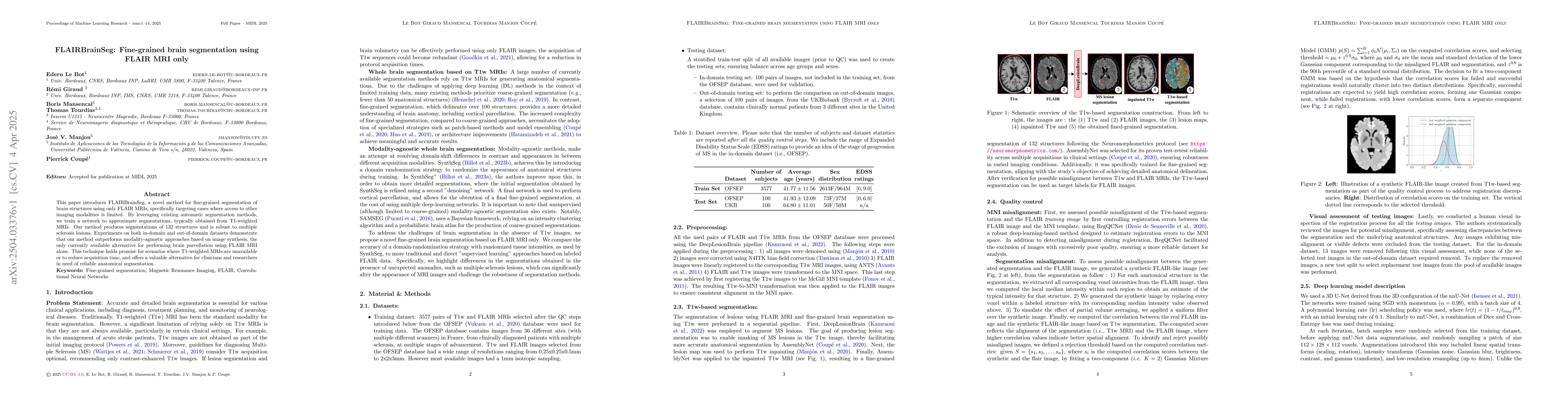
This research introduces FLAIRBrainSeg, a novel framework for fine-grained segmentation of FLAIR MRI scans, specifically targeting cases where T1-weighted MRIs are unavailable. The method leverages existing automatic segmentation methods and trains a network to approximate segmentations typically obtained from T1-weighted MRIs, targeting 132 brain structures with robustness to multiple sclerosis lesions.
Discussion 0