Publication
Metrics
AI Quick Summary
This paper proposes a semi-automatic analysis pipeline for Flash X-ray diffraction imaging (FXI) to convert 2D diffraction patterns into 3D electron densities, including hit finding, pattern classification, 3D reconstruction, and post-analysis with bootstrap methodology. The method was successfully applied to determine the 3D structure of the PR772 virus.
Paper Preview
Abstract
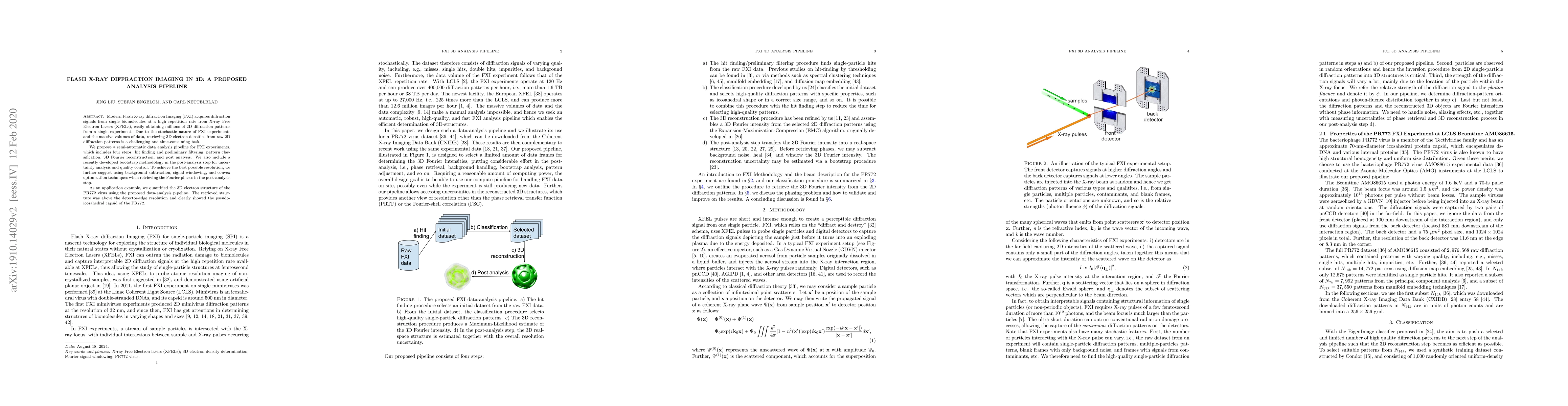
Modern Flash X-ray diffraction Imaging (FXI) acquires diffraction signals from single biomolecules at a high repetition rate from X-ray Free Electron Lasers (XFELs), easily obtaining millions of 2D diffraction patterns from a single experiment. Due to the stochastic nature of FXI experiments and the massive volumes of data, retrieving 3D electron densities from raw 2D diffraction patterns is a challenging and time-consuming task. We propose a semi-automatic data analysis pipeline for FXI experiments, which includes four steps: hit finding and preliminary filtering, pattern classification, 3D Fourier reconstruction, and post analysis. We also include a recently developed bootstrap methodology in the post-analysis step for uncertainty analysis and quality control. To achieve the best possible resolution, we further suggest using background subtraction, signal windowing, and convex optimization techniques when retrieving the Fourier phases in the post-analysis step. As an application example, we quantified the 3D electron structure of the PR772 virus using the proposed data-analysis pipeline. The retrieved structure was above the detector-edge resolution and clearly showed the pseudo-icosahedral capsid of the PR772.
AI Key Findings
Get AI-generated insights about this paper's methodology, results, significance, and more — seven facets brought into focus.
Impact
Paper Details
Authors
PDF Preview
Key Terms
Citation Network
Current paper (gray), citations (green), references (blue)
Display is limited for performance on very large graphs.
Discussion 0